Incidental Findings Associated with Magnetic Resonance Imaging of the Brachial Plexus
Antonia R Perumal, .1, Ugonna Angel Anyamele, .1, Rayna K Bhogal, .1, Gordon McCauley, MBBS1, Irvin Teh, BE MBiomedE PhD1, Grainne Bourke, MB BCh BAO FRCSI FRCS(Plast)2, James J Rankine, MBChB MRCP MRad FRCR MD3 and Ryckie George Wade, MBBS MSc MClinEd MRCS FHEA4, (1)University of Leeds, Leeds, United Kingdom, (2)Leeds Teaching Hospitals Trust, Leeds, United Kingdom, (3)Leeds Teaching Hospitals, Leeds, United Kingdom, (4)Department of Plastic and Reconstructive Surgery, University of Leeds, Leeds, United Kingdom
Title
Incidental Findings Associated with Magnetic Resonance Imaging of the Brachial Plexus
Abstract
Background: The identification and management of incidentalomas is becoming increasingly problematic, particularly in relation to brachial plexus imaging because the prevalence of incidental findings is unknown.
Purpose: To estimate the prevalence of incidental findings in symptomatic patients undergoing MRI of the brachial plexus.
Materials and Methods: This retrospective cohort study includes all children and adults undergoing their first brachial plexus MRI over a 12-year period, in a tertiary care centre in the UK. An incidental finding was any abnormality which was not a direct injury to or disease-process of the brachial plexus. An 'incidentaloma' was defined by the need for further investigation or treatment. Multivariable logistic regression was used to estimate the odds ratio (OR) of an 'incidentaloma'. To estimate which factors were associated with the number of incidental findings, multivariable Poisson regression was used.
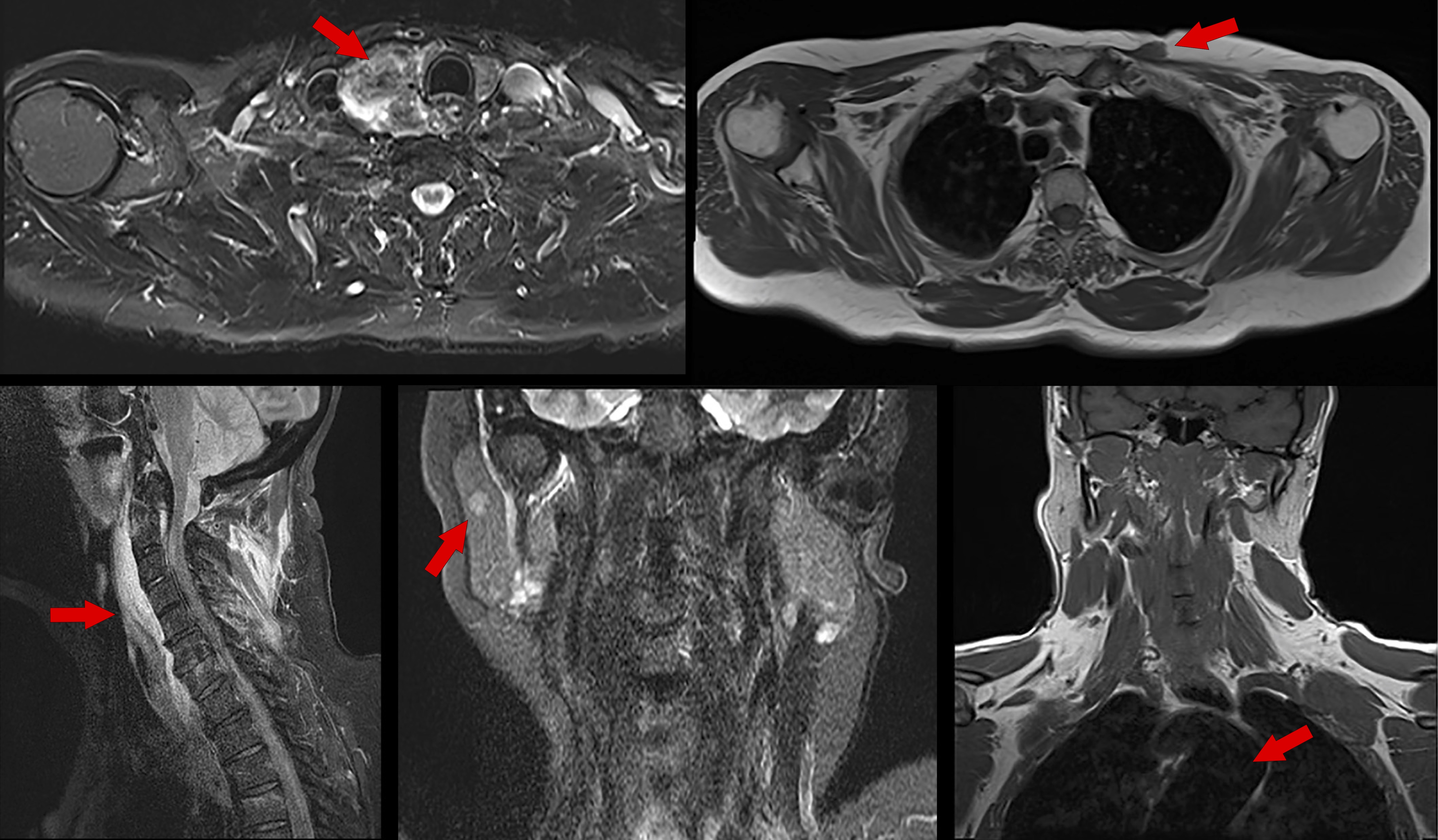
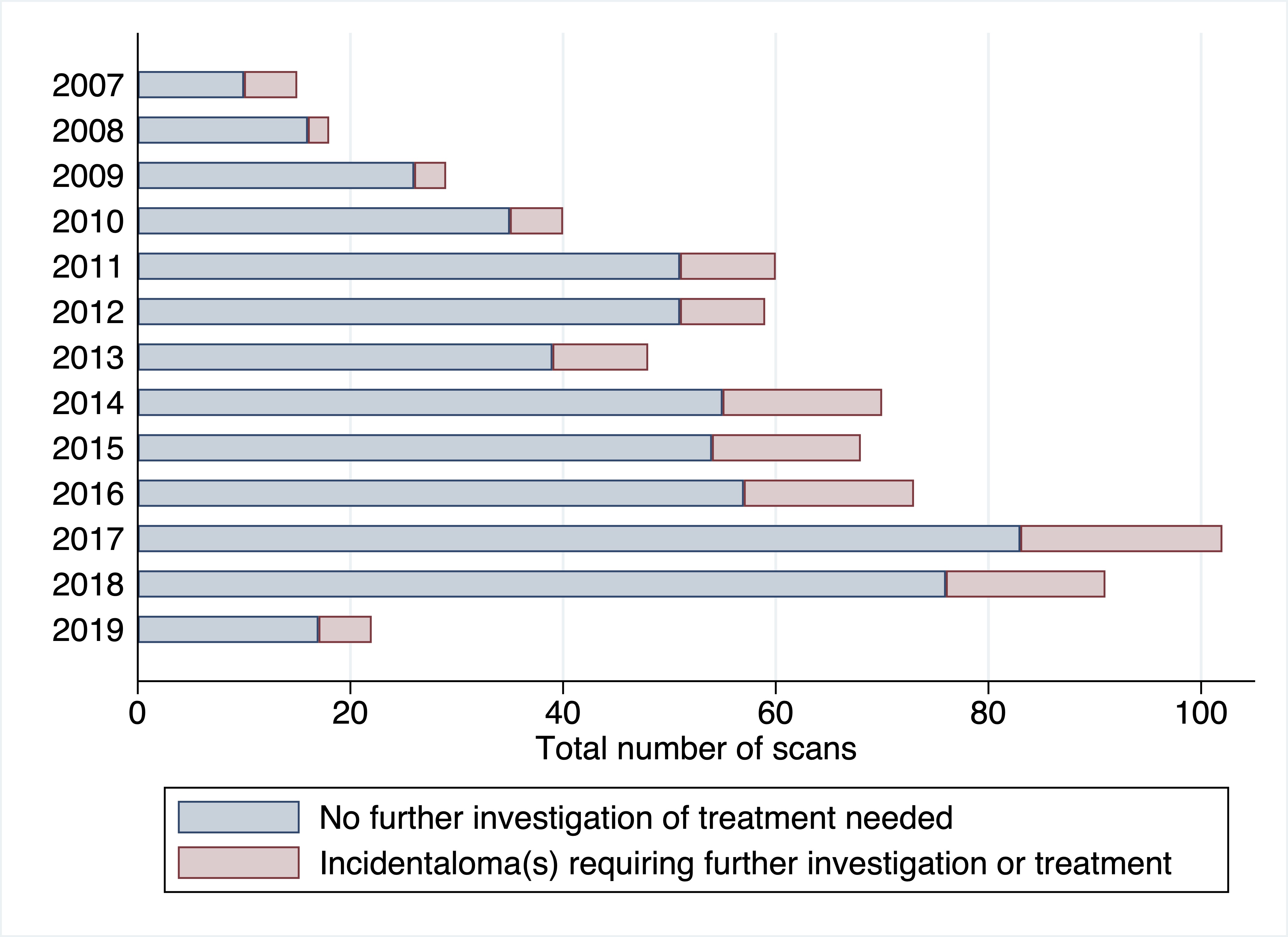
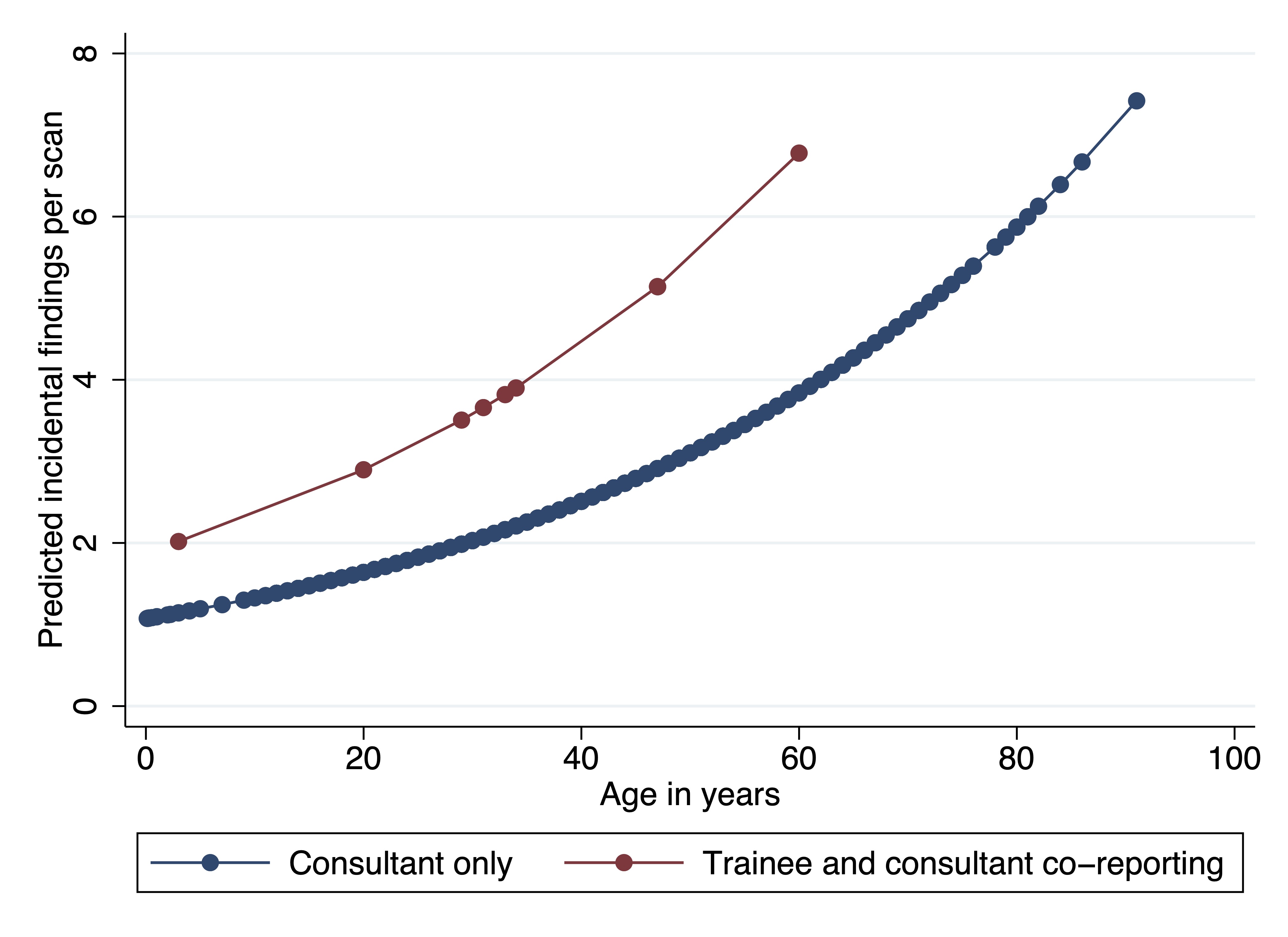
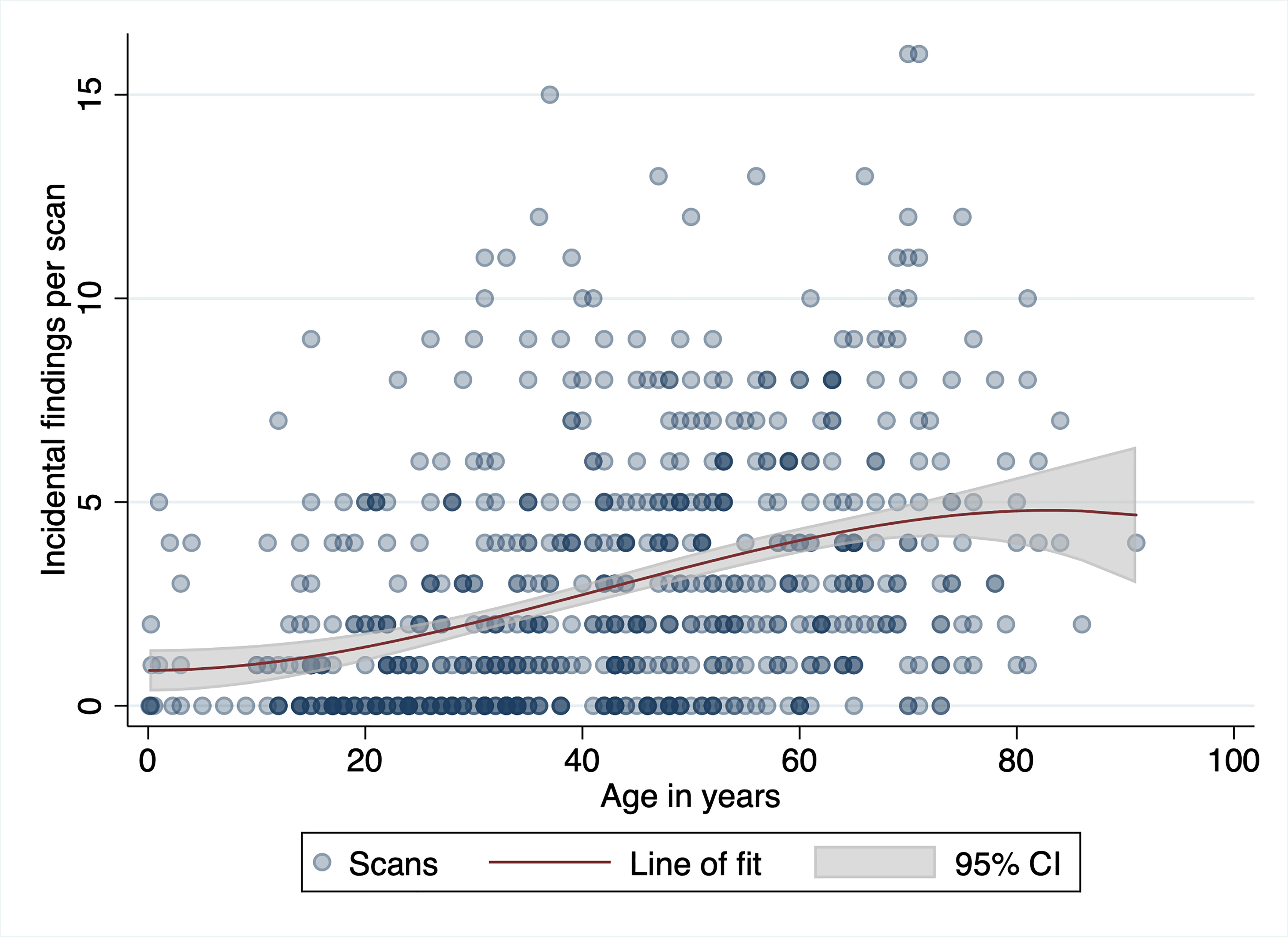
Results: Overall, 502 scans (72%) reported incidental anomalies (Figure 1) of which 125 (18%) required further investigation or treatment. Although the number of MRIs performed per annum increased by 23%, the prevalence of 'incidentalomas' remained static (p=0.766; Figure 2). Musculoskeletal incidental findings were the most prevalent (63%), with a median of 3 per patient. Needing further investigation or treatment was related to the frequency of incidental findings (adjusted OR 1.16 [95% CI 1.08, 1.24]) and tumour identification (adjusted OR 2.86 [95% CI 1.81, 4.53]). The number of incidental findings per scan increased by 36% when trainees co-reported with consultants (Figure 3) and by 2% with every year of life (Figure 4).
Conclusions: The prevalence of clinically important incidental findings on brachial plexus MRI is lower than organ-specific imaging, but still 18% of scans identified an 'incidentaloma' which required further investigation or treatment.
Figure

Figure 2. A stacked bar chart showing the number of times a patient underwent their first MRI of the brachial plexus increased annually by a mean 23%, but the prevalence of incidentalomas remained static.

Figure 3. Scans reported by a trainee and consultant jointly had significantly more incidental anomalies, in patients of all ages.

Figure 4. A scatter plot showing that the number of incidental findings per scan increases with age.

Back to 2021 ePosters
