Alterations in Myelinisation of Painful Nerves, Proximal to the Site of Injury
Elisabeth Maria Brakkee, MD, MSc1, Wim van Hecke, MD1, Erick DeVinney, BS2 and J. Henk Coert, MD, PhD3, (1)UMC Utrecht, Utrecht, Netherlands, (2)Clinical and Translational Sciences, AxoGen Inc, Alachua, FL, (3)University Medical Center Utrecht, Utrecht, Netherlands
Introduction
Damage to motor neurons is often accompanied by cell death in the dorsal root ganglia, which can be prevented by reconnection to a target muscle. After damage to sensory nerve fibers, pain may develop despite of reconstructive surgery. In these patients, usually denervation surgery remains their last treatment option. Previous studies have shown that after nerve lesion, electrophysiological changes develop in the proximal nerve end, which indicates possible irreversible changes. With this study, we aim to demonstrate these proximal structural changes in the sensory nerve after nerve damage on a histopathological level.
Materials& Methods
Surplus of human painful nerve specimen were obtained during routine proximal nerve denervation surgeries. Also, human non-painful nerve specimen were obtained post-mortem from the same anatomical location as where nerve denervation surgeries were performed, serving as a negative control. Signs of demyelinisation and remyelinisation were investigated. Nerve specimens were cut transversely and axon count, Schwann cell count, myelin thickness/G-ratio (measure of maturity of the regenerating nerve) where determined. This study was approved by the Review Committee Biobank.
Results
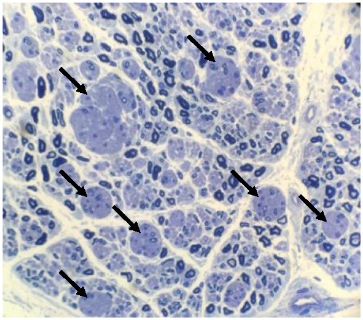
We obtained 6 human painful nerve specimens: radial superficial nerve (RSN) (2x), saphenous (2x), occipital and the palmar branch of the median nerve (more to follow). All the painful nerves showed loss of myelinated fibers, but interestingly not of non-myelinated C-fibers (compared to control sural nerve, post mortem specimen will follow). Two out of six patients show onion bulbs (i.e. concentric layers of Schwann cells surrounding axons), a sign of repetitive demyelinisation and remyelinisation, see figure 1. One patient shows lamination of the myelin sheets. Exact axon count, myelin thickness and G-ratio from the painful and control specimen will be presented.
Conclusions
All painful nerves show loss of myelinated nerve fibers, but no loss of unmyelinated fibers, and thus an altered ratio of myelinated axons/non-myelinated axons. To our knowledge, this histopathological finding of structural changes in the nerve end proximal to the nerve injury are new. Our results concur with previous reports of reduced conduction velocity in the proximal nerve end. We feel that the imbalance between myelinated and non-myelinated nerve fibers may contribute to the development of chronic pain.
Figure 1: Onion bulbs in RSN specimen of a patient with history of denervation surgery in the distal RSN
Back to 2021 ePosters

