
The Role of Corneal Nerves in the Maintenance of Corneal Epithelium
Kiana Tajdaran, MD, MASc1,2, Konstantin Feinberg, PhD3, Seyed Kaveh Mirmoeini, MD, MASc1, Jennifer Zhang, MD, PhD3, Tessa Gordon4 and Gregory H Borschel, MD4, (1)University of Toronto, Toronto, ON, Canada, (2)The Hospital for Sick children, Toronto, ON, Canada, (3)The hospital for sick children, Toronto, ON, Canada, (4)Division of Plastic and Reconstructive Surgery, The Hospital for Sick Children, Toronto, ON, Canada
Background and goal: The cornea is the window through which we see the world and is one of the most densely innervated structures in the body. Besides providing protective sensory input, corneal nerves have been postulated to stimulate limbal stem cells (LSCs), hence governing corneal epithelial maintenance and recovery. Loss of corneal innervation, through injury, diabetes, tumors, infections, and even improper contact lens use, leads to neurotrophic keratopathy (NK), a degenerative corneal disease that is characterized by corneal epithelial breakdown, scarring, and permanent vision loss. Studying the paracrine interactions between corneal nerves and LSCs, and the supporting cell populations inhabiting the LSC niche, would potentially serve as the basis for a pharmacological treatment for NK. Nerve growth factor (NGF) has been suggested to improve epithelial healing in the denervated cornea. However, the mechanism of NGF-induced signaling is not known. The goal of this study is to identify the role of the NGF receptor TrkA in the regulation of LSC.
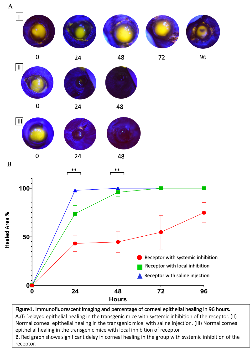
Methods: We chose a transgenic mouse that allows specific pharmacological inhibition of TrkA in vivo. To differentiate between systemic TrkA activity and that locally expressed by LSCs, the mice were treated with the inhibitor, either intraperitoneally or topically, for six days. On day six we removed the corneal epithelium with a 0.5 mm rotating brush. To measure epithelial healing, we performed digital imaging of fluorescein staining daily for four days after injury (Figure 1A). We then harvested the corneas for immunofluorescent and biochemical analyses.
Results: In our mouse model, while the local TrkA inhibition had no detectable effect, the corneal epithelial healing was significantly delayed following systemic inhibition of the receptor. (Figure 1B).
Conclusion: Our results indicate that the activity of TrkA expressed in limbal cell populations, is not directly associated with the regulation of LSC, while the TrkA expressed by other cell populations, such as corneal nerves, is probably involved in stimulation of LSCs and it might affect self-renewal and/or differentiation of the LSCs. These findings inform the development of new and less invasive treatments of NK.
Back to 2021 Abstracts