
Pre-Clinical Investigation of Extracellular Matrix Enhanced Filled Nerve Guidance Conduits Using a Rat Large Distance Sciatic Nerve Defect Model
Alan J Hibbitts, PhD1, Zuzana Koci, PhD2, Simone Kneafsey, BSc2, Adrian Dervan, PhD2, Paige Hinton, MSc2, Leyla Zilic, PhD3, Gang Chen, PhD MD2, Brenton Cavanagh, PhD2, Conor Buckley, PhD3, Simon Archibald, PhD4 and Fergal J O'Brien, PhD2, (1)Royal College of Surgeons Ireland, Dublin, Ireland, (2)Royal College of Surgeons in Ireland, Dublin, Ireland, (3)Trinity College Dublin, Dublin, Ireland, (4)Integra LifeSciences, Princeton, NJ
Introduction:
Biomaterial nerve guidance conduits are an attractive means to repair peripheral nerve injuries. However, healing can be limited due to substrates lacking the necessary pro-repair signalling cues found within the native nerve extracellular matrix (ECM). This often results in poor neurite outgrowth and insufficient blood vessel infiltration. We have developed a collagen-based nerve guidance conduit filled with an aligned microporous internal matrix. This matrix is fabricated from a combination of prominent ECM macromolecules found in the peripheral nerve micro-environment. To comprehensively test these NGCs, they were implanted into a 15 mm rat sciatic nerve defect and regenerative capacity assessed
Materials and Methods:
Outer collagen conduits were filled with an internal matrix of collagen-chondroitin sulphate (Coll-CS). This Coll-CS matrix was further enhanced with fibronectin, laminin 1 and laminin 2 in the ratio (Coll-CS-FibL1L2 1:4:1). Conduits ± FibL1L2 were implanted into a 15 mm sciatic nerve defect in adult Lewis rats. Implants were compared against autograft treated animals for functional recovery and immuno-histochemistry.
Results:
Foot withdrawal and electrophysiology using both conduit types were 85-95% and comparable to autograft treated animals (Figure 1A-C). High levels of neuronal outgrowth and mature myelination were evident (Figure 1 D+E). Angiogenesis was found to be significantly enhanced in Coll-CS-FibL1L2 1:4:1 samples. Outgrowth rates were significantly higher than both non-augmented and autograft samples (Figure 1 F+G).
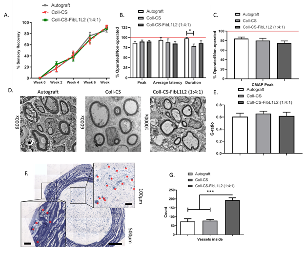
Figure 1. 15 mm rat sciatic nerve recovery at 8 weeks. A) Sensory recovery, B) Compound nerve action potential, C) compound muscle action potential, D) transmission electron microscopy images of mid-graft axons, E) Ratio of the inner to outer radius of myelin sheath (G-ratio), F) mid-graft blood vessel counting and G) quantification
Conclusions:
Addition of ECM macromolecules was found to allow for robust levels of axonal repair in rats. Fibronectin, laminin 1 and laminin 2 combinations significantly increased angiogenesis in the regenerating nerve. This has the potential to allow recovery over even greater distances and is now under investigation in the 30 mm sciatic nerve defect rabbit model.
Acknowledgements:
Co-funded by Science Foundation Ireland and Integra LifeSciences through the AMBER Research Centre (TP27-1846A1)
Back to 2021 Abstracts