
The Effect of Stem Cells and Local Tacrolimus on Neurite Extension
Sara Saffari, MD1, Mana Saffari, MD1, Katelyn Chan, MASc BioSci2, Gregory H Borschel, MD3 and Alexander Y Shin, MD4, (1)Mayo Clinic, Rochester, MN, (2)Division of Plastic and Reconstructive Surgery, The Hospital for Sick Children, Toronto, ON, Canada, (3)Division of Plastic and Reconstructive Surgery, The Hospital for Sick Children, University of Toronto, Toronto, ON, Canada, (4)Microvascular Research Laboratory, Mayo Clinic, Rochester, MN
Background: Application of mesenchymal stem cells (MSCs) or tacrolimus (FK506), an FDA approved immunosuppressant, to nerve grafts has been a topic of interest to enhance peripheral nerve regeneration. The aim of this study was to investigate the combined effect of MSCs and local delivery of FK506 on nerve regeneration when applied to nerve autografts and decellularized nerve allografts.
Materials & Methods: A three-dimensional (3D) in vitro compartmented cell culture system, validated by Tajdaran et al (2019), consisted of rat neonatal dorsal root ganglion (DRG) adjacent to rat nerve autograft or decellularized allograft. This model was used to evaluate regenerating neurites from the DRG into the peripheral nerve scaffold. Nerve autografts and decellularized allografts were augmented with (i) dynamic undifferentiated MSC seeding, (ii) local application of FK506 (100 ng/mL) or (iii) both (N=9/group). Local application was ensured by isolating the central system (i.e. DRG side) from the peripheral system (i.e. nerve graft side), where treatment was applied. After 48-hours of incubation, DRG-nerve graft constructs were collected, fixed, sectioned and stained against neurofilament-160 to measure neurite extension. CD90 staining was used to confirm stem cell characterization.
Results: All grafts treated with MSCs confirmed CD90 expression. Compared to untreated autografts, neurite extension in autografts treated with FK506 and autografts treated with MSCs and FK506 combined were found superior (P<0.001 and P=0.0001, respectively), and comparable to autografts treated with MSCs (P=0.12). Compared to untreated allografts, allografts treated with FK506, and allografts treated with MSCs and FK506 were found superior (P<0.001 and P=0.0001, respectively), and allografts treated with MSCs were found comparable (P=0.09). All autograft groups were found superior compared to their respective allograft treatment group (P<0.05). Solely allografts receiving combined treatment were found superior to untreated autografts (P<0.05).
Conclusions: MSCs or FK506 treatment improved neurite outgrowth and when combined, this resulted in significant synergistic neurite extension in both autografts and allografts in comparable patterns.
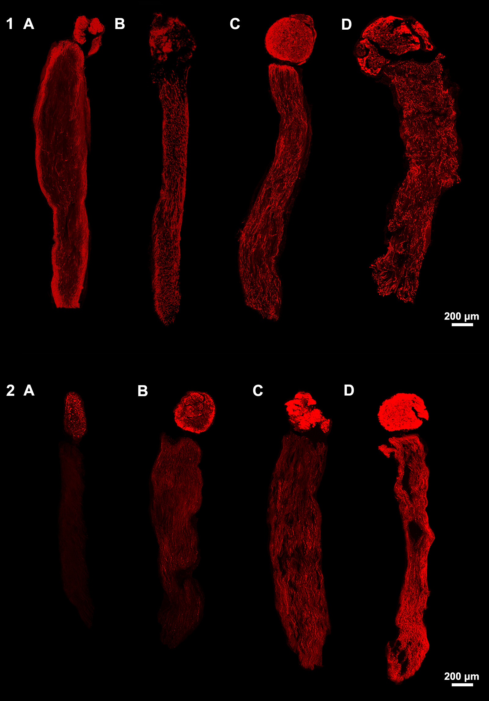
Figure 1. Longitudinal sections of DRG-nerve graft constructs stained against neurofilament-160. Growing neurites in autograft (1A-D) and allograft (2A-D) groups are visualized after application of no treatment (A), seeding with MSCs (B), 100 ng/mL FK506 (C), or MSCs and 100 ng/mL FK506 combined (D). Neurites grew from the DRG into the nerve graft, respectively from the circular top to bottom.
Back to 2021 Abstracts