
Monitoring Regrowth of Injured Peripheral Nerves Using Diffusion Tensor Imaging
Patrick Assi, M.D., Michael Pridmore, M.A., Salam Kassis, M.D., Angel Farinas, M.D., Richard Dortch, PhD and Wesley P. Thayer, M.D., PhD, Vanderbilt University Medical Center, Nashville, TN
Introduction:
Over 17,500 traumatic peripheral nerve injuries (TPNI) to the extremities occur annually in the United States. This study seeks to develop a novel strategy for nerve recovery monitoring based on Diffusion Tensor Imaging (DTI) using Diffusion-Weighted Magnetic Resonance Imaging (DW-MRI). We hypothesize that data acquired via DTI will improve our ability to monitor and predict nerve regrowth following surgical repair of cut or severe crush injuries.
Materials and methods:
DW-MRI studies were performed on 13 patients using a 3.0 T Philips Achieva magnetic resonance scanner. We compared injured median nerves to their correspondent ulnar nerves. One set of data was acquired after initial repair for all 13 patients. Longitudinal data over two timepoints were completed for 3 patients after repair. Qualitative comparison was done using tractography. Quantitative analysis of nerve regrowth was performed using multiple DTI parameters such as: fractional anisotropy (FA), radial diffusivity (RD), axial diffusivity (AD), and mean diffusivity (MD).
Results:
Qualitative comparison on DTI shows axonal regrowth beyond the coaptation site. With a FA closer to 1 correlating with diffusion occurring in a single axis, the quantitative analysis of the data shows that the difference in FA between injured and unaffected nerves is predominantly related to an increased in Radial Diffusivity (RD) which translates into an additional radial regrowth of nerve ending instead of a sole axial growth paralleling the axonal direction (Figure 1). An increase in RD has been correlated historically in the literature with an increase in size of the extracellular compartment surrounding axons. FA improved progressively with time in patients 1 and 2 suggesting a regrowth tendency toward a single axis. With patient 3, we noticed a decrease in FA in the second time period after surgery, but some improvement in FA was witnessed with additional recovery time (Figure 2).
Conclusion:
Early results indicate that DTI using DW-MRI can aid in tracking nerve recovery and potentially guide clinical management either toward or away from surgical re-intervention. The objective of this study is to evaluate the ability of DTI to monitor and to predict nerve regrowth following crush or repaired cut nerve injuries.
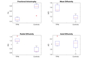
Figure 1
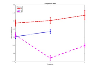
Figure 2
Back to 2020 Abstracts