
The Effect of Crush on Perfusion & Expression of MMP-9 in Rat Sciatic Nerve
David M Brogan, MD, MSc1, Jason M Wever, MS2, Martin I Boyer, MD2, Tony Y Lee, BS2, Samuel Achilefu, Ph.D.2 and Christopher Dy, MD MPH FACS1, (1)Washington University School of Medicine, St. Louis, MO, (2)Washington University in St. Louis, St. Louis, MO
Introduction: Currently available techniques to evaluate axonotmetic nerve injuries are imperfect, making it difficult to localize the zone of injury and severity of injury. Development of intra-operative techniques to better assess these parameters would guide treatment and prognosis. Increased neural vascularity has been found to correlate with chronic nerve compression, but it is unknown whether there is a similar response after acute traumatic nerve injury.
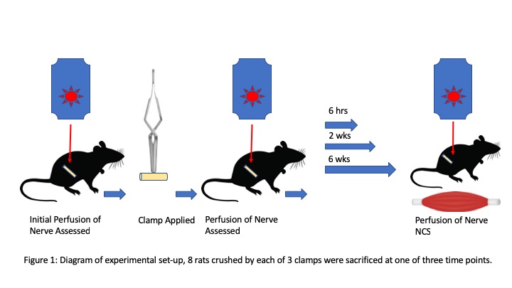
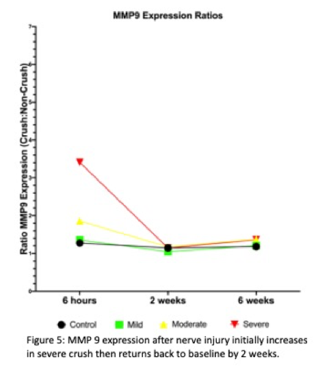
Methods: To address this, we utilized laser doppler flowmetry to assess changes in flux of a rat sciatic nerve at varying time points after a crush injury (Figure 1). Eight rats in each group underwent an initial survival surgery and baseline imaging of red blood cell flux with a laser doppler flowmeter. A graded crush model was created utilizing three distinct vascular clamps with differing degrees of applied force. One clamp per group was applied to the sciatic nerve for 30 seconds. Baseline flux was also measured in the contralateral sciatic nerve to use as a control. After recovering for varying time periods (6 hours, 2 weeks, 6 weeks), laser doppler flow measurements were obtained again at non-survival surgery along with nerve conduction studies and maximal tetanic force measurements. ELISA was used to assay for MMP-9 expression in the control and crushed nerves.
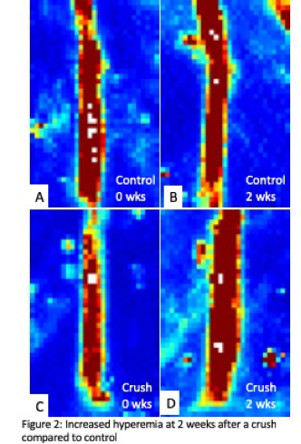
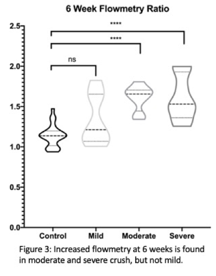
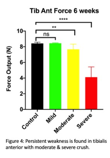
Results: 72 rats underwent the above procedure with varying degrees of crush. Persistent hyperemia was noted in the zone of injury compared to baseline at 2 weeks (Figures 2 & 3) and 6 weeks with incomplete nerve recovery (as evidenced by tibialis anterior muscle weakness) (Figure 4). Gastrocnemius weakness resolved in all animals by 6 weeks and latency of the compound nerve action potential (CNAP) returned to normal. CNAP amplitudes remained diminished when compared to baseline at 6 weeks in all groups. Greater elevation of MMP-9 at 6 hours corresponded to poorer functional recovery of tibialis anterior muscle force, but MMP-9 was not elevated at 2 weeks or 6 weeks (Figure 5). Proximal to the zone of injury, initial hyperemia appeared but then resolved by 6 weeks, suggesting a possible link between location of nerve injury and location of increased blood flow.
Conclusion: These findings suggest that nerve hyperemia and MMP-9 expression may be markers of severe crush injury and resolution of hyperemia may correlate with nerve recovery from trauma. CNAP amplitudes fail to recover to baseline, even at 6 weeks, and may be a more sensitive measure of persistent nerve dysfunction.
Back to 2020 Abstracts