
Neuronal Plasticity: Daily High Electrical Activity Of Cat Hindlimb Motor Nerves And Silencing Of Their Activity By Spinal Isolation and Deafferentation, Decrease And Increase Their Size, Respectively
Tessa Gordon, PhD, Neurosurgery/Spine Center, Division of Plastic and Reconstructive Surgery, The Hospital for Sick Children, Toronto, ON, Canada
Introduction: The effects of neuromuscular activity on nerve caliber are conflicting due to the wide variability in levels of neuromuscular activity from study to study which, in turn, vary with the models of hyper- and hypo-activities employed (reviewed by Gordon & Pattullo Exerc. Sport Sci Rev 21,331-302. 1993). In this study, we addressed the hypotheses that 1) high levels of daily activity of 50%, typical of motoneurons innervating slow motor units, reduce the size of their motor nerves and 2) activities of <1% of daily activity of motoneurons, typical of those of the largest fast glycolytic (FG) motor units (MUs), increase motor nerve size.
Methods: In adult cats, 1) the nerve to medial gastrocnemius (MG) muscle was maximally stimulated with chronically implanted cuff or intramuscular wire electrodes for daily electrical stimulation (ES) at 20Hz in a 50% duty cycle -3 minutes on and 3 minutes off. Conduction velocities (CVs) of single motor nerves were recorded in control cats and chronically for up to 240 days in experimental cats in a final experiment under deep anesthesia, and 2) the spinal cord was transected at L5 and the L5-L7 and S1-S3 dorsal roots transected bilaterally proximal to the root ganglia to isolate and silence motoneurons to MG and soleus muscles. Three weeks and 8 months later, the MG and soleus nerves (n) were removed and post-fixed in osmium tetroxide for measurements of fiber diameters (Dn) and comparison with those from age-matched cats.
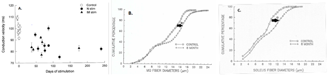
Results: 1) The mean MG nerve CVs declined as a function of time after 50% ES, to a plateau that equaled that of isolated slow nerves and was significantly different from control CVs (p<0.01) (Fig. 1A). 2) The cumulative distributions of the Dn of the silenced MG and the soleus motor nerves were shifted to the right with significant increase (Kolmogorov-Smirnov test, p<0.01) in the size of the motor nerve fibers (Fig, 1B, C), the Ds of the 1a sensory fibers <10µm not changing.
Conclusions: Motor nerves display the same plasticity as the muscles in their adaptation to levels of neuromuscular activity with the size of the nerves decreasing with high levels of activity of the slow MUs and increasing with low or absence activities levels in the fast MUs. This neuronal plasticity is consistent with the known recruitment order of MUs from the smallest most active to the largest and least active FG MUs during movement.
Fig
Back to 2020 Abstracts