
Wrapping Nerves with Conduits Results in Worse Histologic and Functional Outcomes
David M Brogan, MD, MSc1, Christopher Dy, MD MPH FACS1, Jason M Wever, MS2, Dana Rioux-Forker, MD3 and Fraser J. Leversedge, MD4, (1)Washington University School of Medicine, St. Louis, MO, (2)Washington University in St. Louis, St. Louis, MO, (3)University of Missouri, Columbia, MO, (4)Department of Orthopaedic Surgery, DUKE UNIVERSITY, Durham, NC
Introduction: The concept of utilizing a nerve conduit for augmentation of a primary nerve repair has been advocated as a method to prevent neural scarring and decrease adhesions. Despite clinical use, little is known about the effects of a nerve conduit wrapped around a primary repair. To better understand this, we investigated the histologic and functional effects of use of a nerve conduit wrapped around a rat sciatic nerve repair without tension.
Methods: Twenty Lewis rats were divided into two groups of 10 rats each. In each group, unilateral sciatic nerve transection and repair was performed, with the opposite limb utilized as a matched control. In Group 1, direct repair alone was performed, and in Group 2, this repair was augmented with a porcine submucosa conduit wrapped around the repair site. Sciatic functional index was measured at 6 weeks with walking track analysis in both groups. Non-survival surgeries were then performed in all animals to harvest both the experimental and control nerves to measure histomorphometric parameters of recovery. Histomorphometric parameters assessed included total # of neurons, nerve fiber density, nerve fiber width, G-ratio, and % debris. Unpaired t-test was used to compare outcomes between the two groups.
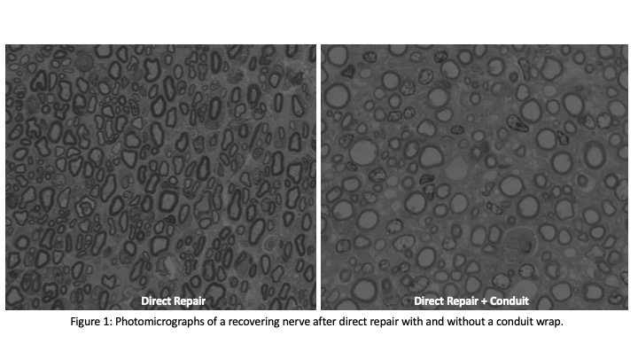
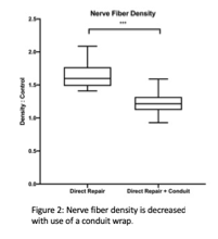
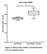
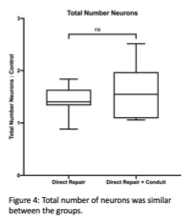
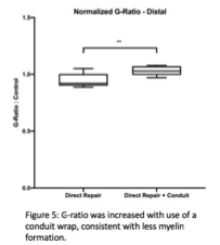
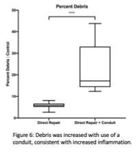
Results: Representative photomicrographs from the second experiment, in which sharply transected nerves were repaired directly with or without a conduit wrap, are seen in Figure 1. The density of nerve fibers was significantly lower in the group with the conduit wrap (p<0.001) (Figure 2) while the fiber width was significantly higher (p<0.001) (Figure 3). The total number of neurons in each did not differ (p=0.52) (Figure 4), but the G-ratio was significantly different between the groups, with the conduit wrap group having a higher G-ratio (p=0.002) consistent with less myelin regeneration (Figure 5). The normalized % debris was significantly higher (p < 0.001) (Figure 6) and the SFI significantly worse (p < 0.001) when a conduit wrap was applied (Figure 7).
Conclusions: Utilization of a conduit wrap after a direct nerve repair was associated with worse remyelination, increased debris, as well as decreased fiber density and worse functional outcomes. More investigations should be performed before routinely recommending the use of a conduit nerve wrap after a primary nerve repair.
Level of Evidence: Level I

Back to 2020 Abstracts