
Developing a Fraction Anisotropy Cutoff Value to Diagnose Recovery in Peripheral Nerve Injury
Angel F. Farinas, MD; Isaac V Manzanera Esteve, PhD; Nancy Cardwell, BS; Alonda Pollins, MLI; Richard D. Dortch, PhD; Mark D. Does, PhD; Wesley P. Thayer, MD, PhD; Galen Perdikis, MD
Vanderbilt University, Nashville, TN
BACKGROUND: Failed peripheral nerve repair can be a devastating condition if intervention is not performed in a timely fashion. Currently, the best way to monitor surgical outcomes is clinical evaluation and nerve conduction studies. Diffuse Tensor Imaging has showed promissory findings to monitor nerve outgrowth. Fractional anisotropy has been correlated with axonal integrity in Central Nervous system. We wanted to create a cutoff value that will be able to classify effectively recovered and non-recovered nerves after repair.
METHODS: 63 Sprague Dawley rats were examined prospectively, divided in three groups: sham, crush and cut/repair. The subjects were evaluated with behavioral tests: Sciatic Function Index and the Foot Fault Asymmetry score; to assess clinical improvement. Nerves were harvested in different time points (1, 2, 4 & 12 weeks). Using a 7T MRI the nerves were scanned. Different parameters were obtained: Axial diffusivity (AD), Radial Diffusivity (RD), Mean Diffusivity(MD) , Fractional Anisotropy and the remaining eigenvalues ( λ1, λ2, λ3). Tractography, a tridimensional axonal representation was elaborated from the DTI parameters.
RESULTS: Radial Diffusivity vs Axial Diffusivity is linearly correlated with FA values of all nerves examined. At 4th week contrast is more evident between recovered and non-recovered subjects based on clinical assessment. Slopes for λ2/ λ1 and λ3/ λ1 were calculated (0.515 ±0.004, 0.328 ± 0.002 respectively) at this time period. Cut off values are obtained using Support Vector Machine (SVM) and Bootstrap technique. A fractional anisotropy cut off value of 0.512 ± 0.002 was obtained. This value was able to classify accurately nerve regeneration at 12 weeks, that correlated with our clinical data and tractography. (Figure 1)
CONCLUSION: A new elaborated fractional anisotropy cutoff value can determine nerve regeneration independently of clinical evaluation, in a prompt and non-invasive way.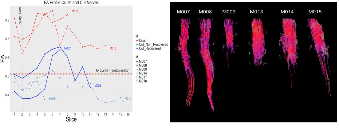
Figure 1. On the left, new FA cutoff value classifying nerve recovery at 12 weeks. On right, tractrogrphy of different subjects in the cut/repair group, correlating with graph on the left.
Back to 2019 Absteracts