
Using DTI MRI to Predict Peripheral Nerve Recovery in a Rat Sciatic Nerve Model
Marlieke Nussenbaum, MD1; Angel Farinas, MD1; Isaac Mazanera Esteve, PhD2; Alonda Pollins, MLI2; Nancy Cardwell, BS2; Richard Dortch, PhD2; Wesley Thayer, MD, PhD3; (1)Vanderbilt University Medical Center, Nashville, TN, (2)Vanderbilt University, Nashville, TN, (3)Department of Plastic Surgery, Vanderbilt University Medical Center, Nashville, TN
Introduction
Peripheral nerve transection is a common injury, with single cut nerve lacerations accounting for over 60% of peripheral nerve surgical interventions in recent studies. For recovery to occur in these patients, axons must grow from the site of repair to the target tissues, a length of up to a meter in humans. Currently, clinical examination and electrodiagnostic tests are the standard practice for peripheral nerve assessment; however neither can rapidly predict whether function will be regained. By the time a diagnosis is made, revisional surgery may not be a viable option due to the onset of irreversible muscle atrophy. Diffusion tensor imaging (DTI) is an MRI sequence that leverages the anisotropic diffusion of water molecules through axons to aid in visualization of neural tracts. DTI parameters, such as fractional anisotropy (FA) and mean diffusivity (MD) are able to quantify microstructural characteristics of nerves, such as axon density and myelin thickness.
Methods
Forty eight female Sprague-Dawley rats were divided into two groups and a sciatic nerve injury model was used. Half of the rats underwent complete nerve transection and immediate microsurgical repair; the other half underwent dissection and exposure of the nerve with no nerve injury (control). Behavioral testing (foot fault and sciatic function index) was performed weekly and the nerves were harvested at 4 different time points: 1 week, 2 weeks, 4 weeks, and 12 weeks, and sent for DTI MRI and subsequent toluidine blue staining for axon counts.
Results
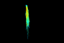
The mean fractional anisotropy of the imaged nerves were calculated for each time point in both the region proximal and distal to the site of injury and repair (Figure 1), with the proximal region showing trends very similar to the control group while the distal region shows a slow increase which mimics results of behavioral testing. Representative fiber tracking based on MRI parameters is shown in Figure 2.
Conclusion
DTI MRI parameters show promise in imaging peripheral nerve injury as well as tracking regain of function following repair.
Figure 1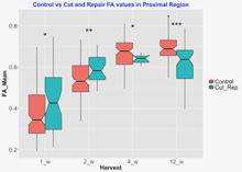
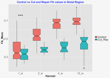
Figure 2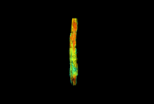
Back to 2018 Program