
Targeted Muscle Reinnervation in the Leg: An Anatomic Study
Megan Fracol, MD1; Lindsay Janes, MD1; Jason H Ko, MD2; Gregory A. Dumanian, MD3; (1)Northwestern University, Chicago, IL, (2)Division of Plastic and Reconstructive Surgery, Northwestern University, Chicago, IL, (3)Division of Plastic Surgery, Northwestern University, Chicago, IL
Introduction:
Targeted muscle reinnervation (TMR) is a surgical technique to re-route the ends of cut nerves to re-innervate small motor nerves of nearby muscles, with the goal of reducing neuroma pain and/or improving prosthesis function. Anatomical roadmaps for TMR have been established in the upper extremity and thigh, but not for the knee and calf.
Materials and Methods:
The major branch points (MBPs) of motor nerves and the motor entry points (MEPs) to muscles of the leg were dissected in five cadaver specimens. Leg length was defined as distance from lateral femoral condyle to lateral malleolus. The distances from the lateral femoral condyle to MBPs and MEPs were measured and these locations were recorded as a percentage of leg length. This information was used to identify targets for TMR in the leg and create a technical roadmap.
Results:
The extensor digitorum longus was identified as the best target in the anterior compartment, with an average of 3 MEPs and 1.3 MBs. All cadavers contained a MEP within 50-70% leg length and MB within 20-45% leg length. The peroneus longus was identified as the best target in the lateral compartment, with an average of 5.8 MEPs and 2 MBs. All cadavers contained multiple MEP within 20-40% leg length and a MB within 20-30% leg length. The gastrocnemius was identified as the best target in the superficial posterior compartment, with an average of 4.4 MEPs per muscle head and 1.6 MBs per muscle head. All cadavers contained multiple MEPs within 10-30% leg length and a MB within 0-10% leg length. The flexor digitorum longus was identified as the best target in the deep posterior compartment, with an average of 6 MEPs and 1.7 MBs. All cadavers contained multiple MEPs within 40-70% leg length and a MB within 20-40% leg length. See figure for example of lateral compartment dissection, green arrows demonstrating MEPs to peroneus longus, blue arrows demonstrating MEPs to peroneus brevis, red arrow demonstrating common peroneal nerve bifurcation, yellow arrows demonstrating superficial peroneal nerve sensory branch, white background demonstrating MBs.
Conclusions:
TMR is technically feasible in the leg with identifiable superior targets in each compartment. This cadaveric study provides a roadmap for incision placement and identification of motor nerve targets when performing TMR in the leg.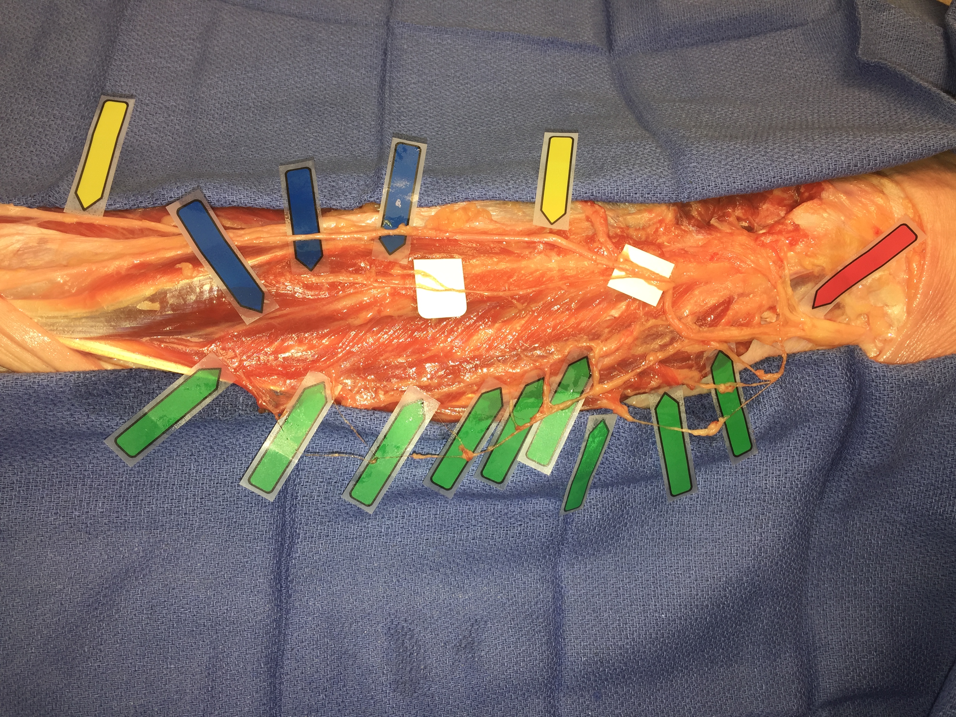
Back to 2018 Program