
Dermal Sensory Interfaces to Restore Graded Sensory Feedback from Prostheses
Andrej Nedic, MSE1; Daniel Ursu, PhD2; Stephen WP Kemp, PhD2; Paul S. Cederna, MD3; Melanie G. Urbanchek, PhD1; (1)Department of Surgery, Section of Plastic Surgery, University of Michigan, Ann Arbor, MI, (2)University of Michigan, Ann Arbor, MI, (3)Section of Plastic Surgery, University of Michigan, Ann Arbor, MI
INTRODUCTION: To provide optimal functional restoration following limb loss, neuroprostheses will require sensory feedback. Dermal Sensory Interfaces (DSIs) are de-epithelialized skin wrapped around the distal end of transected residual sensory nerves. DSIs transmit electrical pulses to generate afferent sensory nerve action potentials. Our purpose was to characterize reinnervated DSI electrical pulse transduction into afferent compound sensory nerve action potentials (CSNAPs) for restoring graded sensory perception.
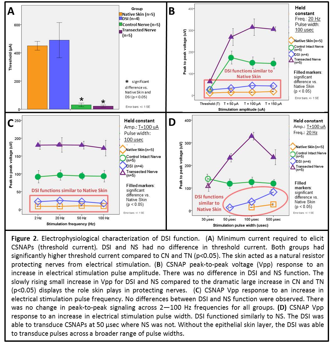
MATERIALS & METHODS: Fischer-344 male rats were used. Control groups were: Native Skin (NS, n=5) and Control Nerve (CN, n=5). Experimental groups were: Transected Nerve (TN, n=5) and DSI (n=4). Sural nerves in TN and DSI rats were transected mid-gastrocnemius muscle. For the DSI group, the transected sural nerve was wrapped with a de-epithelialized glabrous skin graft (Fig. 1). Rats recovered for 2 months. Electrical stimulation was then applied on: ankle skin (NS), the sural nerve (CN and TN), or the DSI. CSNAP peak-to-peak voltage (Vpp) and percent of stimuli transduced (signal sensitivity) were recorded proximally from the sural nerve.
RESULTS: All DSIs were well revascularized with reinnervation by the sural nerve. Importantly, for DSIs, increasing stimulation amplitude from threshold (T) to T+150 µA resulted in an anticipated increase in Vpp similar to the control NS group (Fig. 2). The slowly rising slope represents the role skin plays in protecting the well-being of nerves and allows for signal modulation when compared with the dramatically steep rise in Vpp of CN and TN (p<0.05). There were no significant differences in Vpp when varying stimulation frequency for all groups. For DSIs, there was a predictable increase in Vpp with pulse width increase (13.5±7.3 µV at 30 µsec to 81.7±36.0 µV at 500 µsec; R2=0.524) with no difference from NS at 100 to 500 µsec. Most notably DSIs could transduce electrical pulses below 100 µsec where NS could not. Electrical stimulation applied to DSIs reliably elicited CSNAPs with signal sensitivity ?93%.
CONCLUSIONS: DSIs successfully transduced electrical stimulation to CSNAPs that replicate native intact skin. This study suggests that modulating stimulation pulse amplitude or pulse width may provide graded sensory feedback from neuroprosthetic devices in patients with limb loss. 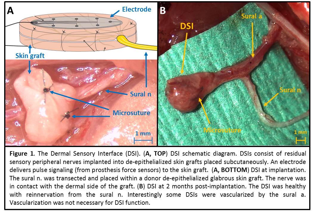
Back to 2018 Program