
Development of Targeted Muscle Re-innervation model in Hind-limb Amputated Rats
Rohit Garg, MD; Safak Uygur, MD; Joanna Cwykiel, MS; Maria Siemionow, MD PhD DSc; University of Illinois at Chicago, Chicago, IL
INTRODUCTION
Symptomatic neuromas can be extremely painful and are often present after traumatic limb amputations. Myriad of surgical techniques have been described with conflicting results and lack of a definitive surgical management. Targeted muscle re-innervation (TMR), originally used to enable control of upper limb prostheses, has been shown to be effective in resolution of post-amputation neuroma pain in clinical studies. The purpose of this study was to develop a TMR model in hind-limb amputated rats for future assessment of neuroma prevention.
MATERIALS and METHODS
Ten hind limbs from Five Sprague Dawley cadaver rats (250-300 g) were used for this study. Sciatic nerve, main branches of sciatic nerve (common peroneal, tibial, saphenous), motor branches from sciatic nerve to biceps femoris and cauda femoris, gluteal nerve and its motor branches to the semimembranosus and biceps femoris and femoral nerve were dissected to look for consistent nerve anatomy that can be used for TMR in the rat hind limb amputation model. Transfemoral amputation was performed and two types of coaptations were made:
1. Common peroneal and motor branch to biceps femoris(branch of gluteal nerve)
2. Tibial nerve and motor branch to semimembranosus(branch of gluteal nerve)
RESULTS
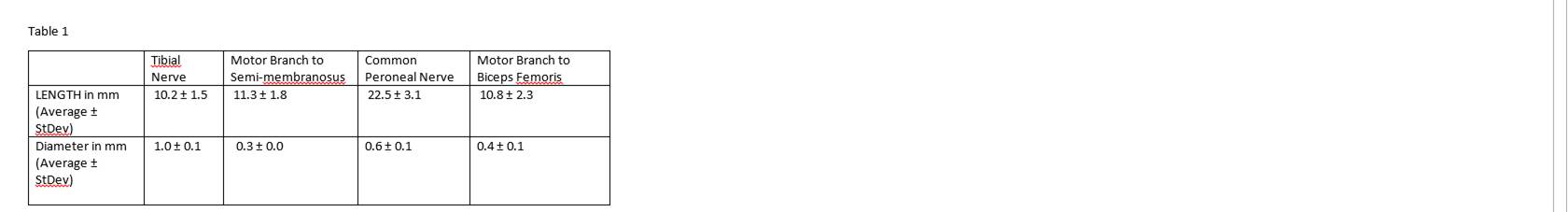
The total surgical time for the dissection, amputation and coaptation of nerves was approximately 90 minutes. A total of 100 nerves were dissected in 10 rat hind limbs. Anatomy of the dissected nerves was consistent. Hind-limb amputations were performed without damaging the target muscles and nerves. Nerve lengths were sufficient for coaptation without any tension. The average lengths and diameters of nerves utilized for neurotization are summarized in Table 1.
CONCLUSIONS
To the best of our knowledge this is the first TMR model in hind limb amputated rats. Rat models are more readily available and cost effective. The coaptation between common peroneal and motor branch to biceps femoris and tibial nerve and motor branch to semimembranosus was found to be the most feasible method for TMR considering the limitations in size and diameter mismatch between other donor and recipient nerves. This model will allow for mechanical, EMG and histological analysis for future assessment of neuroma prevention.
Back to 2018 Program