The Spiral Structure of Peripheral Nerve and its Clinical Relevance
Antonio Merolli, MD, FBSE1; Marco De Spirito, PhD2; Joachim Kohn, PhD FBSE1
1Rutgers University, Piscataway, NJ, , 2UniversitÓ Cattolica, Rome, Italy
Introduction: In 1779 Fontana described an ondulating arrangement of nerve fibers in peripheral nerves. This arrangement produces the optical phenomenon of the bands of Fontana. In 1968 Sunderland described a plexiform arrangement for the fascicles in peripheral nerves. This showed how the single nerve fiber can change its course along the nerve. We analyzed how this spiral structure gives a contribution in explaining the clinical efficacy of nerve regeneration and repair by artificial nerve-guides (conduits). These devices are clinically effective despite the fact that they do not provide any actual fiber-matching clue.
Materials and Methods: Microdissections were performed in twenty sciatic nerves of Wistar Rats and high-speed digital video-recording was performed prior to sacrifice. Standard optical and confocal microscopy were performed on explanted nerves.
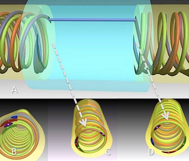
Results: The wave, as early described by Fontana, is actually a bi-dimensional representation of a three-dimensional spiral. The three-dimensional structure of the peripheral nerve at rest can be described as an assembly of spirals (or coils). This spiral arrangement is functional in responding to a stretching force eventually applied to the nerve. It may provide as well, however, a statistical match for fibers regenerating inside an artificial nerve-guide (conduit). Fibers in the proximal stump will be evenly distributed all around the nerve cross-section. This radial spreading enhances the probability that at least some fiber will reconnect with its proper functional distal end. As the specific signal may be transmitted by more than one fiber due to a redundancy in transmission, a limited number of successful matches may provide a sufficient clinical recovery.
Conclusions: The spiral arrangement of nerve fibers provides an in-built mechanism so that a limited number of functional connections, established by the use of an artificial nerve-guide (conduit), may result in an adequate clinical recovery.
Figure 1: (A) In a gap-lesion repaired by a cylindrical artificial nerve-guide, fibers spiralling at a certain pitch will evenly spread all around the circumference (B). This means that for any bundle of fibers in the proximal stump (C), there will be always a computable probability that some of them will match the correct target (D)
Back to 2017 ePoster Listing
