
Back to 2017 Annual Meeting Program
InVivo Optical Microscopy to Investigate Revascularization and Regeneration Following Peripheral Nerve Repair
Jeena M Easow, MD; Isabel Chico-Calero, PhD, DVM; Ahhyun S Nam, PhD; Mark A Randolph, MAS; Benjamin J Vakoc, PhD; Robert W Redmond, PhD; Jonathan M Winograd, MD
Massachusetts General Hospital, Boston, MA
Introduction: Re-establishment of blood supply to the injured nerve has been postulated as a necessary component to neural recovery. The process by which this occurs and the impact of graft revascularization on the regenerative process remains poorly understood.
The objective of this study was to image the process of neural revascularization in conjunction with regeneration and remyelination using optical frequency domain imaging (OFDI). OFDI is a non-invasive, optical imaging modality that uses low-power infrared light to visualize tissue microstructure. Functional extensions of OFDI, such as angiography and polarization-sensitive platforms are used to simultaneously acquire data regarding tissue vascularization and myelination with spatial registration of tissue microanatomy.
Methods: The rodent sciatic nerve model was utilized for the following experimental groups: 1) crush injury, 2) repair with 5 mm nerve autograft, and 3) repair with 5 mm acellular nerve allograft. OFDI was used to perform intravital imaging at baseline, post-operative days 7, 14, and 28. Angiographic-OFDI was used to detect microvascular networks in the surrounding nerve bed and epineurium. Polarization Sensitive-OFDI was used to assess axonal myelination as it correlated to nerve birefringence. Auto-fluorescence detection was used to measure axonal regeneration in these thy1-GFP rats. Experimental groups were then compared regarding blood vessel formation, axonal myelination, axonal regeneration, and histomorphometry.
Results: In vivo imaging showed a robust neovascularization stemming from the surrounding tissues to the site of injury at day 7 and subsequent remodeling as evidenced by the presence of epineural vessels by day 28 (Image 1). On post-operative day 7, notable loss of fluorescence (axonal) and birefringence (myelin) at the site of injury and distally had occurred. Marginal recovery was visualized by day 28; crush>autograft>allograft. Correlating histomorphometry is in progress.
Conclusions: OFDI has sufficient spatial resolution to permit clear visualization of neural microvasculature and allows for quantitative measurements of neural birefringence enabling the examination of spatial and temporal reestablishment of blood supply and myelination following nerve repair. Based on preliminary imaging results, there is a significant increase in vascularity originating from the surrounding tissue with a development of epineural vessels in crush, autograft and even allograft groups, albeit slowest in the allograft group. This non-invasive technology provides greater insight into the role of neovascularization in peripheral nerve regeneration, particularly in the setting of autografts and allografts.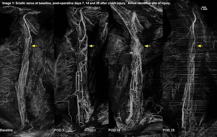
Back to 2017 Annual Meeting Program