
Back to 2017 Annual Meeting Program
Macaques Implanted with Regenerative Peripheral Nerve Interfaces (RPNIs) Control Prosthesis Finger Movements
Stephen WP Kemp, PhD; Melanie G Urbanchek, PhD; Zachary T Irwin, PhD; Philip Vu, MSE; Shoshana L Woo, MD; Ian C Sando, MD; Jana D Moon, BS; Cynthia A Chestek, PhD; Paul S Cederna, MD
University of Michigan, Ann Arbor, MI
Introduction: Regenerative Peripheral Nerve Interfaces (RPNIs) are promising for interfacing human intentions to myoelectric prostheses. Rat studies led to proof of RPNI long-term function and high signal to noise ratio with no adverse biological effects. However, true voluntary fine control of fingers and hand prostheses would be more convincing with macaque implanted RPNIs.
Methods: Two macaques had Regenerative Peripheral Nerve Interfaces (RPNIs) implanted (n=3/macaque) in the forearm. The RPNI consists of a free muscle graft implanted on the end of a transected nerve fascicle. Intramuscular EMG electrodes were implanted in each RPNI. Macaques were trained to perform index finger movements to acquire virtual targets on a computer screen. Finger position was recorded via a flex sensor on the index finger.
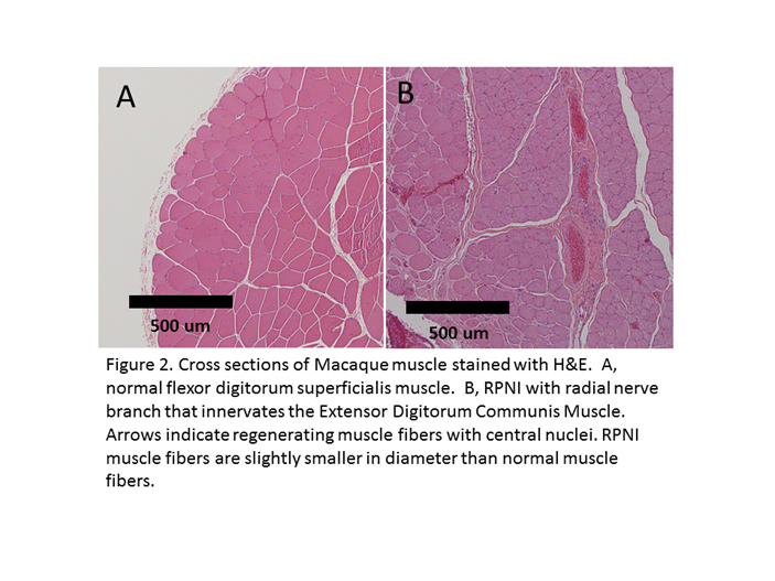
Results: At harvest RPNIs were well vascularized but smaller in size than when implanted (Fig 1). For the continuous EMG decode using 10-fold cross-validation, the resulting predicted finger position had a, correlation coefficient ?=0.82 between predicted and true finger positions. The EMG decode correctly classified 97.7% of movements (out of 261 total movements). RPNI muscle fibers were continuing to regenerate after implantation for 1 year (Fig 2).
Conclusions: Macaques voluntarily controlled virtual finger movements with signals transferred through implanted RPNIs.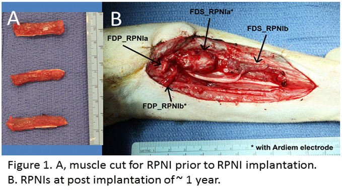
Back to 2017 Annual Meeting Program