
Back to 2017 Annual Meeting Program
A Rat Model Of Corneal Denervation And Neurotization Using the Thy1-GFP+ Rat
Joseph Catapano, MD1; Jennifer J Zhang, MD, PhD2; Gregory H. Borschel, MD2
1University of Toronto, Toronto, ON, Canada, 2The Hospital for Sick Children, Toronto, ON, Canada
Introduction: Corneal neurotization significantly improves corneal sensation in patients with corneal anesthesia, using nerve grafts and donors from the contralateral face to reinnervate the denervated cornea. Important questions remain, including whether donor sensory nerves, foreign to the cornea, maintain corneal clarity and how the cornea regulates nerve ingrowth after neurotization. In this study, we developed an experimental animal model of corneal neurotization where experimental factors can be manipulated and tissue harvested, in order to investigate these questions.
Methods: Our model of corneal neurotization was designed using the thy1-GFP+ rat, which expresses green fluorescent protein (GFP) in axons. First, stereotactic surgery was performed to electrosurgically ablate the ophthalmic nerve (V1), which innervates the cornea, using a 21G insulated needle. Multiple stereotactic variables, including the stereotactic coordinates, number of lesion sites, and the amount and duration of energy stimulus were investigated (n = 75). The cornea was examined 1, 2 and 4 weeks later for effective denervation with whole-mount confocal microscopy. Once corneal denervation was established, a second group of rats (n = 12) was used to establish a method of corneal neurotization. Common peroneal (CP) and sural nerve grafts were coapted to the contralateral inferior orbital nerve (ION) and then sutured to the corneal limbus for corneal neurotization. Corneal reinnervation was examined and compared to non-neurotized controls 2 and 4 weeks later with the same microscopic method.
Results: Optimal stereotactic corneal denervation was achieved by ablating two locations of the intracranial ophthalmic nerve at the coordinates 2.1, 1.5, 11 and 2.3, 0, 11 mm with electrosurgical parameters of a 390 kHz sinusoid waveform and 3 W administered for 30 s at each coordinate. The cornea remained denervated for 2 weeks with minimal reinnervation at 4 weeks (Fig 1B). ION reinnervation of the cornea through CP and sural autografts occurred within 4 weeks of denervation (Fig 1C).
Conclusions: We have established a rat model of corneal neurotization by optimizing stereotactic parameters to ablate the corneal innervation intracranially and successfully neurotizing the corneal using nerve grafts from the ION. This provides an excellent model to study corneal neurotization and in turn, optimize clinical corneal reinnervation. 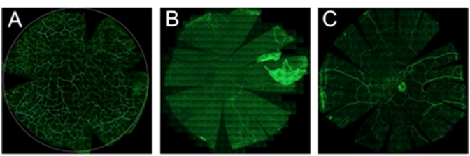
Figure 1. Thy1-GFP+ axons in the uninjured cornea (A) are absent 4 weeks after denervation (B). Significant corneal reinnervation occurs 4 weeks after corneal neurotization (C).
Back to 2017 Annual Meeting Program