
Back to 2017 Annual Meeting Program
Proximal versus Distal Nerve Transfer for Biceps Reinnervation – A Comparative Study in the Rat Model for Brachial Plexus Injury
Johnny Chuieng - Yi Lu, MD; Aleksandra McGrath, MD, PhD; Frank Fang, MD; Tommy Nai-Jen Chang, MD; David Chwei-Chin Chaung, MD
Chang Gung Memorial Hospital, Kweishan, Taiwan
Background: Debate persists between proponents of proximal and distal nerve transfers for brachial plexus reconstruction. The best role for each method has not yet been defined. Here, we use a rat model to compare proximal versus distal nerve reconstruction strategies.
Methods: Male Sprague-Dawley rats were used. C6 spinal nerve with nerve graft (proximal transfer model, n=42) and 50% of ulnar nerve (distal transfer model, n=42) were used as the donor nerves (Figure 1). The target for reinnervation was the musculocutaneous nerve (MCN). Outcomes were recorded at 4, 8, 12, and 16 weeks. Outcome parameters were grooming test, biceps muscle weight, compound muscle action potentials, tetanic contraction force, and axonal morphology of the MCN.
Results: The axonal morphology revealed no significant difference between groups. Ultimately, the proximal transfer model produced superior outcomes at 16 weeks compared to the distal transfer model (Figure 2 to 5). The proximal transfer group's time interval analysis showed a peak in axonal counts at 12 weeks with trend of improvement in all functional and physiologic parameters across all time points. In contrast, the distal transfer group reached its peak performance at 8 weeks and plateaued from 8 to 16 weeks. Also, its axonal counts were highest at the initial 4 week time point with some axonal loss occurring thereafter.
Conclusion: Proximal nerve transfer outcomes are superior to distal nerve transfer in our experimental model. The distal nerve transfer group reaches an early peak performance plateau, but the proximal nerve transfer group progressively improves and eventually surpasses the distal nerve transfer group.
Figure 1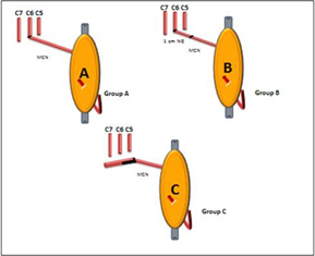
Figure 2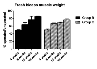
Figure 3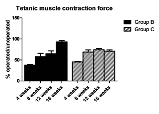
Figure 4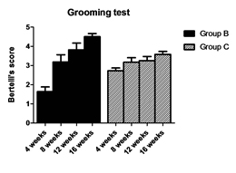
Figure 5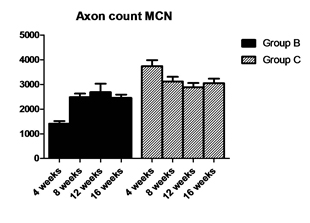
Back to 2017 Annual Meeting Program