
Back to 2017 Annual Meeting Program
Experimental Validation of a New Rabbit Model of Brachial Plexus Injury
Kathleen Kollitz, MD; Patricia F. Friedrich, BA; Allen T. Bishop, MD; Alexander Y. Shin, MD
Mayo Clinic, Rochester, MN
Introduction: Though the rat have sciatic nerve has frequently been used to model peripheral nerve injury, it is not suitable for study of the brachial plexus. We describe a true brachial plexus injury model in the rabbit upper extremity. We have found the biceps to be reliably innervated by the middle trunk (formed by the C6 and C7 nerve roots). We aim to demonstrate both long-term biceps paralysis when this trunk is severed, and recovery after nerve repair. We hypothesized that middle trunk repair would result in significantly larger muscle cross-sectional area (CSA), biceps isometric tetanic force (ITF) , wet muscle weight (WMW) and compound muscle action potentials (CMAP)than those left unrepaired.
Methods: 22 rabbits underwent bilateral ultrasound measurement of biceps CSA prior to unilateral surgical division of the middle trunk. Five rabbits were randomized to the "no repair" group. The remaining 17 rabbits underwent direct coaptation. CSA was measured at 4 weeks, 8 weeks and 12 weeks post-operatively. At 12 weeks, rabbits were sacrificed and bilateral CMAP and ITF were measured. Biceps WMW was recorded, and its motor nerve analyzed for total nerve area, myelinated area, axon area, axon count and N-ratio (myelinated area/total nerve area). Results from the operative side were expressed a percentage of the non-operated side, and groups compared across outcomes.
Results: By 8 weeks post-operatively, the CSA of the no repair biceps measured 64.4% of the non-operated side while the repair group measured 78.0% (p=0.05). By 12 weeks CSA fell to 59.3% for the unrepaired group and increased to 89.1% for the repair group (p=0.008, Figure 1). At sacrifice, biceps WMW was significantly higher in the repair group (65.8%±19.4%) as compared to the no repair group (52.0%±9.6%, p=0.047). The repair group rabbits exhibited significantly higher CMAP (23.3%±29.3%, p=.0048) and ITF (25.6%±37.4%, p=0.012) as compared to the no repair group, which exhibited no recovery.
Conclusions: The middle trunk is the main source of innervation to the rabbit biceps muscle. This model can be successfully used for evaluation of reinnervation of the biceps muscle. Ultrasound can be used as a non-invasive measure to monitor recovery of biceps muscle function over time. Based on our findings, a survival time longer than 12 weeks is recommended for future investigations. 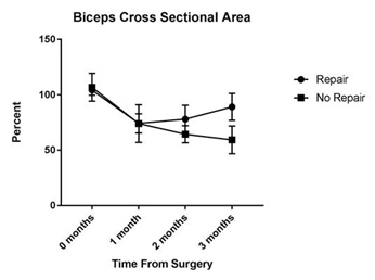
Back to 2017 Annual Meeting Program