
Back to 2015 Annual Meeting Program
Selective Nerve Root Transection Produces a Permanent, Partial Nerve Injury Model
Louis H. Poppler, MD; Matthew D. Wood, PhD; Daniel A. Hunter; Lauren Prange; Susan E. Mackinnon, MD; Amy M. Moore, MD
Department of Surgery, Division of Plastic and Reconstructive Surgery, Washington University School of Medicine, St. Louis, MO
Introduction: This study's goal was to develop a partial, non-regenerative nerve injury model that results in permanently reduced muscle force to allow the study of therapeutics to improve motor function. Reduction of the motorneuron pool to 20% normal values is required to produce a measureable loss of muscle force in rats. We hypothesized that transection of one or more of the L4-6 nerve roots would cause a permanent measurable reduction in muscle force.
Methods: Eighty rats were randomized into four groups (n=20) that underwent variations of nerve root transections. Group I and II had the L4 or L5 nerve root transected respectively, and a silicone cap placed proximally to prevent regeneration. Group III had both L4&5 roots and group IV had the L4&6 roots transected and capped. Retrograde labeling of the tibial and peroneal nerves (n=12 per group) was performed at 3 weeks. Normal values were established in a sham surgery group. Tibial and peroneal nerves were harvested for histomorphometry at 3 and 12 weeks to evaluate the presence of myelinated axons. Muscle force testing (n=8 per group) was conducted to provide functional data corroborating the reduced counts. Neurman-Keuls post-hoc analysis was performed.
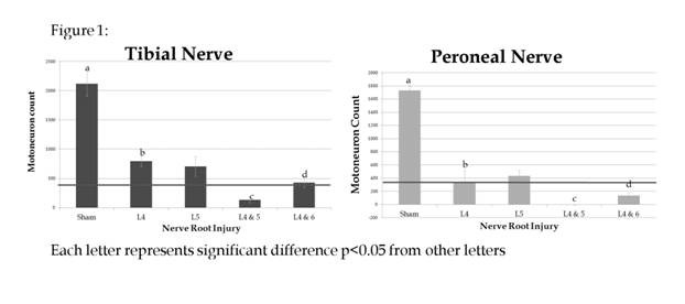
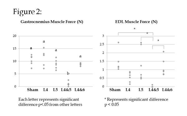
Results: In group III (L4&5-cut), the tibial mean motoneuron count was 6% of control, and peroneal nerve had no motoneurons. In group IV (L4&6-cut) mean motoneuron counts in the tibial and peroneal were 20% and 8% respectively (Figure 1). Large myelinated axon counts confirmed these results. In group III, the muscle mass of gastrocnemeus and extensor digitorum longus (EDL) were significantly reduced v. sham (p<0.05). In group III, mean gastrocnemeus force was to 1.1N (9.6% of sham), a statistically significant reduction. EDL force was 0.0 N in group III, confirming motoneuron and myelinated axon counts (Figure 2). No significant reduction in muscle force was observed in other nerve injury groups.
Summary Points:
Ģ This partial nerve injury model produces reproducible and stable decrease in muscle force.
Ģ Unlike previously described partial nerve injury models, this novel model avoids the rat's innate neuroregenerative capability and produces a stable platform from which to evaluate therapeutics to increase motor function.
Back to 2015 Annual Meeting Program