
Back to 2015 Annual Meeting Program
Histological Comparison of Tibial and Peroneal Nerves Following Stretch Injury
Nicholas Schraut, MD1; Sharon Walton, MD1; Jad Bou Monsef, MD1; Susan Shott, PhD2; Anthony Serici, BS2; Lioubov Soulii, MD1; Farid Amirouche, PhD1; Mark Gonzalez, MD1; James Kerns, PhD1
1Orthopaedic Surgery, University of Illinois Chicago, Chicago, IL; 2Rush University, Chicago, IL
Introduction: There are over 1 million total joint arthroplasties done annually in the United States, with nerve injury occurring in 0.5-2.0% of cases. Most of these injuries occur to the peroneal nerve; its sequelae can cause significant morbidity. Our goal in this this study was to recreate a nerve stretch injury that would be encountered during TJA; to investigate histological differences in the peroneal and tibial nerves versus control nerves.
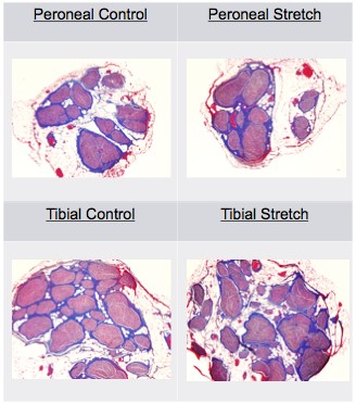
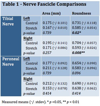
Methods: The peroneal and tibial nerves were harvested bilaterally from 10 fresh humans cadavers. Each nerve was sectioned into 2 samples. The control sample was suspended from a frame with sutures at its native length and placed in Karnovsky's Fixative. The other sample was tied with sutures to a frame and stretched to 20% of its native length and placed in fixative. After 1 week in fixative, each nerve was washed 3 times in 0.1M cacodylate buffer, sectioned, and stained with Masson's trichrome. Using ImageJ software, each nerve was analyzed for its overall and individual fascicular cross sectional areas, and roundness [(4xarea) / (pixdiameter2)] (as roundness approaches zero, the fascicle is more oval). Statistical comparisons were performed using SPSS software with the Friedman test (significance at alpha < 0.05%).
Results: The tibial nerves' fascicles decrease in size upon stretching (Table 1); the peroneal nerve has variable changes in area upon stretch (Figure 1). The tibial nerve significantly decreases in roundness with stretch whereas the peroneal nerve does not show a significant change. The proportion of the epineurial tissue that was connective tissue had a tendency to be greater in the tibial nerves (49.6-55.4%) compared to the peroneal nerves (38.1-41.9%). The tibial nerve has significantly more fascicles (29.5) than the peroneal nerve (13.4)(p < 0.001).
Conclusion: The peroneal nerve is more prone to injury during total knee and total hip arthroplasty than the tibial nerve. Upon stretching, the peroneal and tibial nerves behave differently, which could account for their difference in injury rates. Though both nerves decrease in area when stretched, the tibial nerve changes in its shape from circular to more oblong; the peroneal nerve stays circular. Perhaps the tibial nerve's higher composition of connective tissue or higher fascicle number accounts for these differences and gives the tibial nerve greater protection. The present study provides new insights into differences between the two main nerves, which may help explain why the peroneal nerve is more at risk of injury with stretch.
Back to 2015 Annual Meeting Program