
Back to 2015 Annual Meeting Program
Compression Neuropathy of the Common Peroneal Nerve Due to an Intramuscular Ganglion Cyst
Katelin K. O'Brien, BS; Michael L. Singer, BS; Jessica Korsh, MS; Kristin Aliano, MD; Thomas A. Davenport, MD; Laurence Glickman, MD
Research, Long Island Plastic Surgical Group, PC, Garden City, NY
Introduction: Compression of the common peroneal nerve (CPN) by an extraneural ganglion causing foot drop is a rare entity; the literature reveals few reported cases. A ganglion is rarely painful but associated symptoms can occur due to either cyst rupture or pressure on adjacent structures. This case report may be of value for awareness and differential diagnosis of progressive peroneal palsy. Early diagnosis and decompression can lead to the best possible neurological recovery.
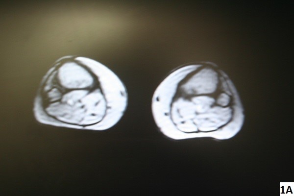
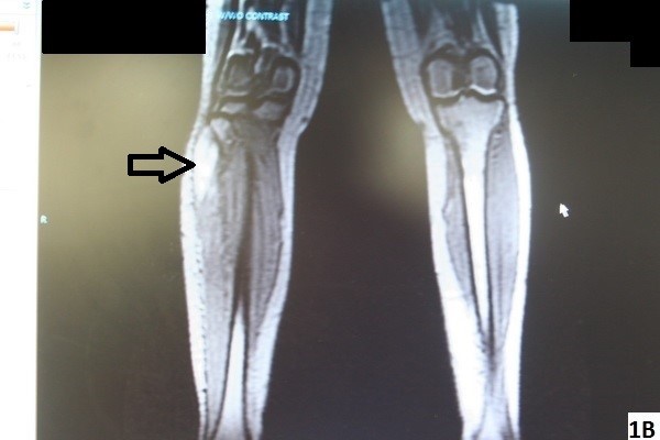
Materials & Methods: A 60-year-old woman presented to the emergency department with pain over the lateral aspect of the right lower extremity, loss of sensation in the first and second web spaces, and an inability to dorsiflex the right foot. The physical exam was remarkable for pain and swelling over the head of the fibula and her history was negative for trauma. MRI revealed a mass overlying the right fibular head with CPN compression. The patient underwent excision of an intramuscular mass in the anterior tibilias muscle and pathology exam of the mass revealed a ganglion cyst.
Results: Five weeks postoperatively, the patient had excellent dorsiflexion of the affected foot and great toe. However, the patient still experienced minimal residual weakness of great toe extension. Deep peroneal nerve sensibility was restored by five weeks.
Conclusion: Ganglionic impingement neuropathy presents more challenges to the physician and may require multiple imaging and operative interventions to diagnose and prevent permanent impairment4. Although compression of the peroneal nerve by an intramuscular ganglion cyst is rare, this entity should be considered in the differential diagnosis for the cause of foot drop.
Figure 1A & 1B. MRI imaging revealing a lesion consistent with a benign cyst.
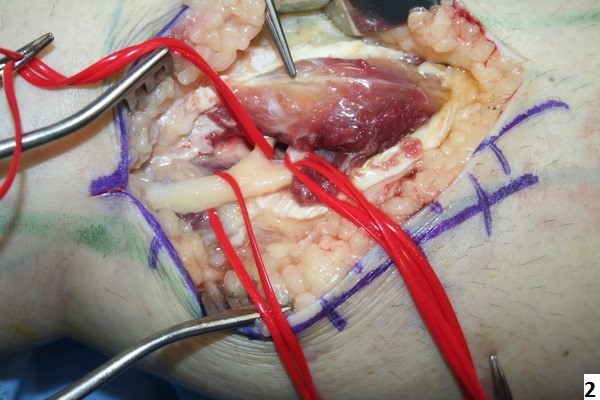
Figure 2. Intraoperative image demonstrating the location of the ganglion within the anterior tibilias muscle compressing the CPN.
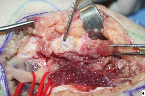
Figure 3. Exposure of Intramuscular Ganglion Cyst.
Back to 2015 Annual Meeting Program