
Back to 2015 Annual Meeting Program
Modelling the Effect of Monopolar Electrical Stimulation on Axonal Activation within a Sensory Regenerative Peripheral Nerve Interface
Xin Zheng, BS1; Cynthia A. Chestek, PhD1; Shoshana L. Woo, MD2; Melanie G. Urbanchek, PhD2; Paul S. Cederna, MD2; Nicholas B. Langhals, PhD2
1Biomedical Engineering, University of Michigan, Ann Arbor, MI; 2Plastic Surgery, University of Michigan, Ann Arbor, MI
Introduction: Current state-of-the-art bio-engineered prosthetic limbs have the potential to restore cutaneous sensation via direct nerve stimulation; however, the mechanical mismatch between the rigid alloplastic electrodes and the soft biologic nerve negatively affects long term nerve health and signal quality. Our laboratory has developed an experimental sensory regenerative peripheral nerve interface (sRPNI) by implanting a residual sensory nerve into a freely grafted piece of muscle. Sensory axons in the sRPNI can then be indirectly stimulated, avoiding trauma induced by direct stimulation. sRPNIs have been viable in rats for four months, with axonal sprouting throughout the muscle and no apparent neuroma formation. Our goals were to model the effect of sRPNI stimulation on axonal activation and quantify the percent neural activation as a function of stimulation location and intensity.
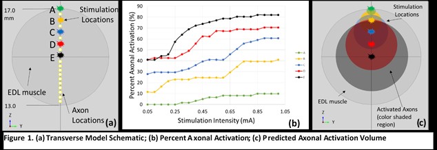
Methods: A finite element model of the sRPNI was developed using COMSOL (a finite element solver). Muscle (ellipsoidal, 335 mm3, conductivity: longitudinal = 0.5, transverse = 0.1 S/m) of the sRPNI was virtually placed in a saline tank (0.9%, 37°C, 2 S/m). Monopolar currents (0.05-1 mA) were injected at throughout the sRPNI. The calculated voltage field for each stimulus was used to compute the internal currents of the axons within MATLAB. These currents were compared to the threshold for sural axon activation previously evaluated in NEURON (a modelling software for neural tissues) and percent axonal activation was calculated.
Results: At stimulation locations within the sRPNI, percent axonal activation was calculated for increasing stimulation intensities (Figure 1). Stimulating at the surface of the sRPNI and the center of the RPNI resulted in 10% and 80% activation at 1 mA, respectively. Threshold axonal activation is observed as low as 250 µA at the surface and 50 µA in the center.
Conclusions: We developed a finite element model of a sRPNI that demonstrates the feasibility of activating restricted populations of axons, thereby permitting the transmission of differential sensations via a prosthesis to its user. The more central the stimulation, the greater the percent axonal activation. This model will help guide the development of our sRPNI stimulation algorithms to restore the sense of touch to prosthetic users, improving not only their function, but also their quality of life.
Back to 2015 Annual Meeting Program