
Back to 2015 Annual Meeting Program
A Novel Immunomodulatory Approach using a Selectively Permeable Nanofiber Wrap to Enhance Nerve Regeneration
Karim A. Sarhane, MD, MSc; Zuhaib Ibrahim, MD; Russel Martin, MD; Kellin Krick, MD; Chris Cashman, MD; Sami H. Tuffaha, MD; Justin M. Broyles, MD; Nijaguna Prasad, MD; Sepehr Tehrani, MD;Christopher Wallner, MD; Mohammad Alrakan, MD; Damon S. Cooney,MD; Ruifa Mi,MD; Ahmet Hoke, MD; WP Andrew Lee, MD; Hai-Quan Mao, MD; Gerald Brandacher, MD
Department of Plastic and Reconstructive Surgery, Johns Hopkins University, Baltimore, MD
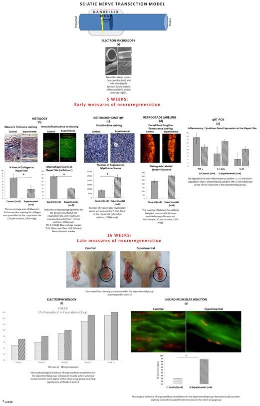
Purpose: Despite great advances in microsurgery, functional outcomes following nerve repair remain suboptimal. Scar formation at the repair site is recognized as a major impediment to regenerating axons. In this regard, an inert barrier around the coaptation site that prevents inflammatory cells infiltration while still allowing the diffusion of nutrients and nerve growth factors holds great potential in promoting nerve regeneration and functional return. In this study, we examined the efficacy of a novel semi-permeable nanofiber construct, prepared from FDA approved biomaterials, to be used as a wrap around the repair site to promote nerve regeneration and functional recovery.
Methods: Nerve wraps comprised of nonwoven electrospun poly (?-caprolactone) nanofibers with pores smaller than 10 µm were synthesized (Fig. 1a). They were wrapped around the repair site in a sciatic transection/repair model in Thy-1 GFP rats. At 5 weeks, their neuro-protective and neuro-regenerative potentials were assessed. At 16 weeks, functional recovery was evaluated.
Results: At 5 weeks, the nanofiber wraps resulted in significantly decreased collagen deposition and inflammation/macrophage invasion at the repair site (Fig. 1b). The total number of myelinated axons was significantly increased (Fig. 1d), and there was a trend towards a higher number of regenerated dorsal root ganglion sensory neurons. Mechanistically, these outcomes were correlated to an up-regulation of the anti-inflammatory cytokine (IL-10) and down-regulation of the pro-inflammatory cytokine (TNF-?) (Fig. 1e). In addition, at 16 weeks, the nerve wrap group showed enhanced functional recovery as demonstrated by electrophysiology (Fig. 1f), gait analysis, neuromuscular junction re-innervation (Fig. 1g), and gastrocnemius muscle weight and histology.
Conclusions: Our results demonstrate favorable outcomes of a novel semi-permeable and clinically translatable nanofiber nerve wrap in protecting the coaptation site and enhancing axonal regeneration through scar-free nerve repair, resulting in optimal functional recovery.

Back to 2015 Annual Meeting Program