
Back to 2014 Annual Meeting Program
Minced Nerve Tissue In Vein Grafts Used As Conduits In Rat Tibial Nerves
Cihan Sahin, MD1, Huseyin Karagoz, MD, PhD1, Yalcin Kulahci, MD, Associate, Pro2, Celalettin Sever, MD1, Dilek Akakin, MD3, Bircan Kolbasi3, Ersin Ulkur, MD1 and Fatih Peker, MD4
1Plastic, Reconstructive & Aesthetic Surgery, GATA Haydarpasa Training Hospital, Istanbul, Turkey, 2Department of Hand and Upper Extremity Surgery, Gulhane Military Medical Academy, Ankara, Turkey, 3Histology and Embryology Department, Marmara University School of Medicine, Istanbul, Turkey, 4F&P Plastic Reconstructive and Aesthetic Surgery Center, Istanbul, Turkey
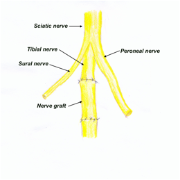
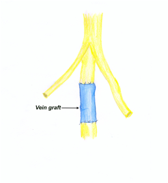
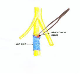
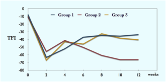
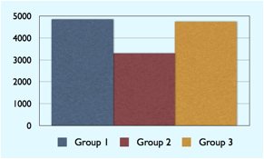
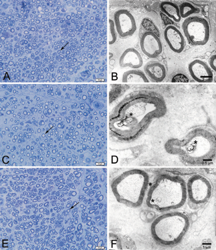
Peripheral nerve injuries are encountered frequently in clinical practice. In nerve repair, an end-to-end suture is the preferable choice of treatment. However, where primary closure is not possible, the defect is to be repaired with a nerve graft. A total of 21 female Wistar rats were used in the study. They were classified into three groups: (I) nerve graft, (II) vein graft, and (III) minced nerve graft. In group I, following exposure of the tibial nerve, a 1cm-long nerve gap was created on the tibial nerve, and the defect was repaired epineurally by using the autogenous nerve. In group II, the 1cm tibial nerve defect was repaired by using an autogenous vein graft. In group III, a 1cm nerve graft divided three equal parts, with one of the nerve parts being minced with micro-scissors and placed in the vein graft lumen. Thereafter, a 1cm tibial nerve defect was repaired by the vein graft filled with minced nerve tissue. The tibial function indices (TFIs) were calculated for functional assessment using the Bain-Mackinnon-Hunter formula. Light and electron microscopic evaluations were performed for morphometric assessment. In addition, the myelinated fibers were counted in all groups. The TFIs of group II were found to be the lowest among all the groups after the 6th week, whereas the TFI of group I was found to be better than the other groups after the 6th week. There was no difference in TFIs between group I and group III. Based on the number of myelinated fibers, there was no statistically significant difference between group I and group III, whereas the difference was significant (p < 0.05) between groups I/III and group II. Presence of peripheric nerves in light microscopic evaluation revealed normal characteristics of myelinated fibers in all groups. The myelinated axon profile was near normal in the nerve graft group in electron microscopic evaluation. However, there were more degenerated axons with disturbed contours and vacuolizations in the vein graft group compared to the minced nerve graft group. We can conclude that using minced nerve tissue in vein grafts as a conduit increases the regeneration of nerves and it may not be caused by donor site morbidity. It can be used in the repair of nerve defects instead of autogenous nerve grafts after further experimental evidence and clinical trials.
 |
 |
| Walking Track Analysis | Myelinated Fibre Count |
Light and electron microscopic photomicrographs of all groups.
Back to 2014 Annual Meeting Program