
Back to 2014 Annual Meeting Program
Development of Regenerative Peripheral Nerve Interfaces for Somatosensory Feedback
John V. Larson, BS, Jana D. Moon, BS, Theodore A. Kung, MD, Melanie G. Urbanchek, PhD, Paul S. Cederna, MD and Nicholas B. Langhals, PhD
Department of Surgery, Section of Plastic Surgery, University of Michigan, Ann Arbor, MI
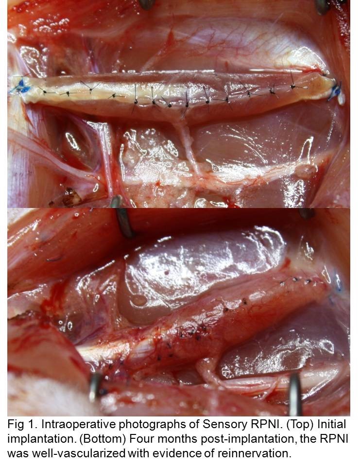
Introduction: Major technological advances have improved the functional capability of bioengineered prosthetic limbs. However, the ability to provide long-term, distinguishable, graded somatosensory feedback from the prosthesis to the patient has yet to be achieved. Our laboratory has developed a prototype sensory neural interface to create and transmit sensory input to the residual limb, providing high-fidelity sensation through advanced neuroprostheses. Methods: Twelve rats underwent sensory RPNI fabrication whereby the left extensor digitorum longus (EDL) muscle was elevated as a free-muscle transfer to the ipsilateral thigh and reinnervated by the transected sural sensory nerve. The entire construct was then encircled with small intestinal submucosa. To demonstrate reinnervation, after a convalescent period of 3 - 4 months, in vivo sensory recordings were obtained from the sensory RPNI while electrically stimulating the sural nerve. RPNIs, sural nerves, and contralateral control EDL muscles and sural nerves were then harvested for mass comparisons. Results: Upon gross examination, RPNIs appeared well vascularized, healthy, and viable (Figure 1). During electrophysiological testing, the average stimulation threshold required for eliciting a compound action potential was 99.6 µA in the 4-month RPNIs and 143.75 µA in the 3-month RPNIs, indicating continued regeneration and reinnervation. Furthermore, the 4 month RPNIs demonstrated an average compound action potential peak-to-peak amplitude of 2.27 mV, compared with 0.68 mV in the 3 month RPNIs. As expected, both groups exhibited similar muscle latencies. Additional EMG data for the 3 and 4 month groups are summarized in Table 1. Direct RPNI electrical stimulation resulted in contraction, confirming muscle health. RPNI muscle mass was up to 75.19% that of control EDL muscle (SD 0.1268). Comparisons of electrophysiological responses and muscle mass indicated that muscle and nerve fiber recovery approached equivalence. Conclusion: These findings demonstrate that freely transferred muscle becomes reinnervated when neurotized by a transected sensory nerve. Furthermore, electrophysiological signal is successfully transmitted between the sensory RPNI and residual nerve. This functional sensory neurotization can be used to provide amputees with the sense of touch. Future work will examine tissue reinnervation through histological analysis of RPNIs as well as afferent neural signal generation through these constructs. 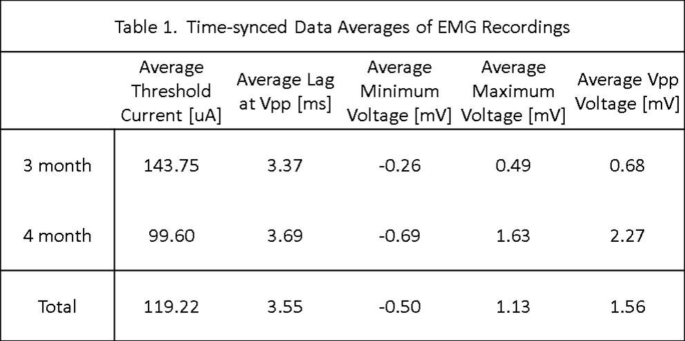
Back to 2014 Annual Meeting Program