
Back to 2014 Annual Meeting Program
Reliability and Validity of Regenerative Peripheral Nerve Interface Function during Voluntary Movement
Andrej Nedic, MSE1, Daniel Ursu, MS2, Jana D. Moon, BS1, Brent Gillespie, PhD2, Nicholas B. Langhals, PhD3, Paul S. Cederna, MD1 and Melanie G. Urbanchek, PhD3
1Plastic & Reconstructive Surgery, University of Michigan, Ann Arbor, MI, 2Mechanical Engineering, University of Michigan, Ann Arbor, MI, 3Department of Surgery, Section of Plastic Surgery, University of Michigan, Ann Arbor, MI
INTRODUCTION
Regenerative Peripheral Nerve Interface (RPNI) devices successfully transduce peripheral nerve action potentials to electrical signals suitable for prosthesis control. Voltage changes are the controlling mechanism and can be observed during electromyography (EMG). RPNI device signaling hasn't been extensively characterized during awake, voluntary movements. Our study: a) characterizes RPNI active to background signal EMG strength and b) defines the reliability and validity of RPNI EMG function during purposeful movements. METHODS
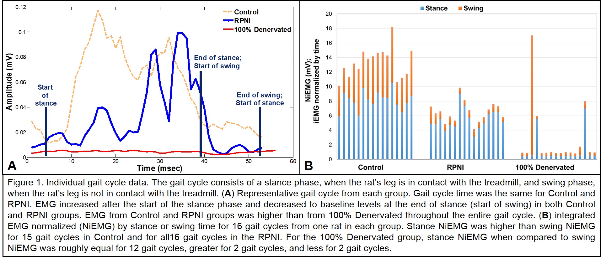
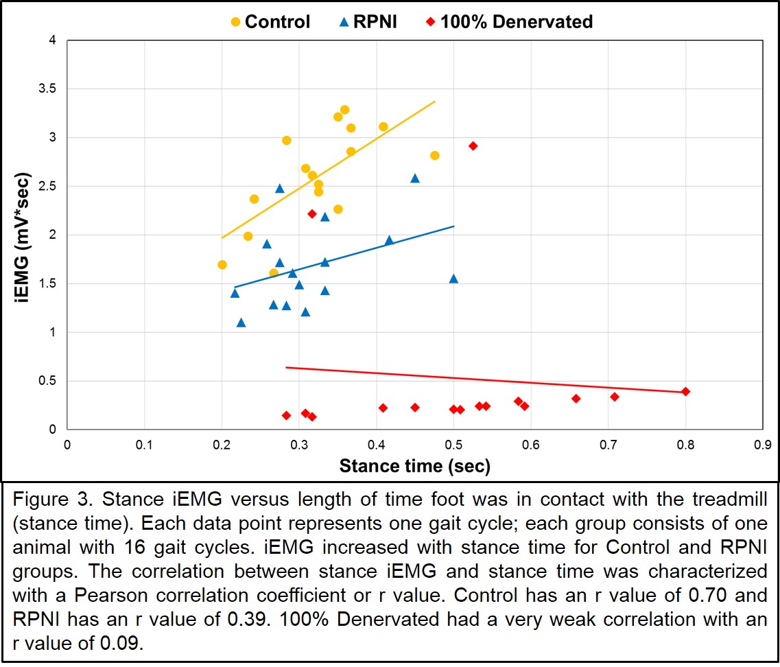
Three groups were formed in rats: Control (n=3), RPNI (n=3), 100% Denervated (n=3). Bipolar electrodes were implanted onto the soleus muscles in each group. For RPNI devices, the soleus muscle was freely grafted to the ipsilateral femur and neurotized by the transected tibial nerve. In the 100% Denervated, the tibial nerve was transected. While walking on a treadmill, rats were video-graphed and raw EMG signals were simultaneously recorded. Video and EMG recordings were synchronized by time for stance, swing, and sit (nonactive) gait phases. Rectified EMG was integrated (iEMG) for each gait phase (Fig. 1). iEMG was normalized (NiEMG) to time for each phase. Data represent 16 gait cycles for each of 3 rats. Correlations were performed between iEMG and stance time to determine reliability. RPNI signaling was validated against Control group signal timing by step phases using Chi Square analysis. RESULTS
We compared EMG signals to background signal strength in all groups during all gait phases (Table 1, Fig. 1). Fidelity of RPNI activity (stance) to background signaling (sit) was 5 to 1, higher than Control signal fidelity (Fig. 2). Significant differences between stance and swing NiEMG activity were confirmed for the Control and RPNI groups. As expected, stance and swing EMG signals were not different for the Denervated group. Correlations between iEMG and stance time for the Control (r=0.7) and RPNI (r=0.4) indicate "good" RPNI signal reliability (Fig. 3). NiEMG signals increased at the start of stance and fell to baseline at the start of swing in both Control and RPNI rat gait cycles (Fig. 1A).These data comparing step cycle to EMG activation accuracy between Control, RPNI, and Denervated groups validated RPNI signaling as purposeful peripheral nerve activity appropriate for meaningful control of prostheses movements (Chi Square; p<0.5). CONCLUSION
RPNI signal fidelity, reliability and validity were examined during voluntary movement. With select filtering, signal fidelity was clear. RPNI signal reliability during the gait stance was "good". RPNI signaling was successfully validated against normal peripheral nerve signaling during walking. 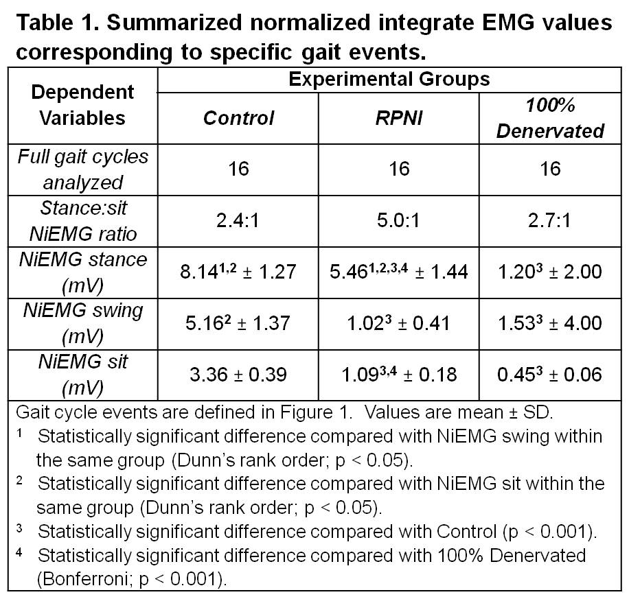

Back to 2014 Annual Meeting Program