
Back to 2014 Annual Meeting Program
Diffusion Tensor Tractography for Assessing Peripheral Nerve Injury and Repair
Richard Boyer1, Kevin Sexton, MD1, Nathaniel Kelm2, David Colton Riley1, R. Bruce Shack, MD1, Mark Does, PhD2 and Wesley Thayer, MD, PhD1
1Department of Plastic Surgery, Vanderbilt University Medical Center, Nashville, TN, 2Department of Biomedical Engineering, Vanderbilt University Institute of Imaging Science, Nashville, TN
Background: Peripheral nerve injury research is highly dependent on animal models and behavioral testing methods, such as sciatic functional index, which are expensive, time-consuming and unreliable. Histologic morphometric analysis and electron microscopy are used for quantitation of axonal regeneration, but these methods cannot be used in-vivo and therefore require animals to be sacrificed at multiple time points. There is limited research on the use of non-invasive imaging for analysis of regeneration after peripheral nerve injuries. Preliminary studies have shown promising results with diffusion tensor tractography (DTT) imaging in a crush model of peripheral nerve injury. In this study, we have applied a similar diffusion-weighted MRI imaging sequence to rat sciatic nerves with controlled degrees of severance injury and microsurgical repair. Methods: Sprague Dawley rats were anesthetized with isoflurane and underwent partial or complete sciatic nerve transection followed by microsurgical epineurial repair. Rats were sacrificed at one hour post-repair and the injured nerves were excised and immersion-fixed with 4% glutaraldehyde/0.5% paraformaldehyde in PBS for 24 hours. Images were acquired using a 2D diffusion-weighted spin echo sequence on a 4.7T 31-cm horizontal bore Agilent DirectDrive MRI scanner. A single 3-mm thick sagittal slice was used to encompass the entire nerve. Diffusion weighted images were reconstructed and fractional anisotropy (FA) maps generated using Diffusion Toolbox 0.6.2. Tractography was performed using the Fiber Assignment by Continuous Tracking (FACT) method with FA threshold of 0.25. TrackVis 0.5.2 was used for fiber tract visualization. Fiber counts were determined by the number of proximally seeded fibers that tracked 1-mm distal to the zone of injury. Results: Four rats were included in each group of partial transection, complete transection and control (no injury). Figure 1 shows diffusion tensor tractography of representative nerves in each group with tracts seeded proximal to the zone of injury. Tract counts in completely transected nerves (1.8 ± 0.92 tracts) were significantly lower than both partially transected (155.8 ± 45.5, p=0.0136) and control nerves (199.7 ± 94.6, p=0.0286). Conclusion: DTT is a promising non-invasive modality for imaging peripheral nerve injury and regeneration. Additional studies will be performed to assess this technique for in-vivo imaging. 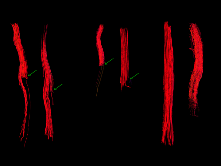 Figure 1: Representative tractography of partially transected (left), completely transected (center) and control (right) nerves harvested immediately following repair. Fiber tracts are shown in red with the proximal nerve at the top. Green arrows indicate zone of injury.
Back to 2014 Annual Meeting Program