
Back to 2014 Annual Meeting Program
A Novel Imaging Technique for Evaluating Axonal Regeneration after Peripheral Nerve Transection in a Rodent Model
Joanna H. Ng, MD1, Yookyung Jung, PhD2, Cameron P. Keating, MD1, Prabhu Senthil-Kumar, MD1, Amanda M. Meppelink, BS1, Mark A. Randolph, MS1, Conor L. Evans, PhD2 and Jonathan M. Winograd, MD1
1Division of Plastic and Reconstructive Surgery, Massachusetts General Hospital, Boston, MA, 2Wellman Center for Photomedicine, Massachusetts General Hospital, Boston, MA
BACKGROUND: Current nerve regeneration outcomes, including walking tracks, electrophysiology, and histology are limited by multiple factors, including interobserver reliability, inaccurate inkprints, short distances and destructive tissue sectioning. Imaging techniques such as confocal microscopy, can provide high resolution images but are limited by natural indices of refraction, light scatter, and suboptimal depth penetration (100-200µm maximum). We sought to resolve this dilemma by utilizing a tissue clearing method, previously applied to the central nervous system, to homogenize the index of refraction and reduce light scatter via chemical replacement of water and lipids. By increasing depth penetration, it is possible to identify individual axons and provide an alternative to traditional immunohistochemical and histomorphometric methods of evaluation. In combination with coherent anti-stokes Raman scattering (CARS) microscopy, a multifaceted image of both myelin sheaths, axons, and the neuromuscular junction can be generated. METHODS: Ten thy-1 transgenic green fluorescent protein (gfp) and wild type rats underwent sciatic nerve transection and repair with standard microsurgery. Nerve restoration was evaluated 2, 4, 7, and 30 days after repair by walking track analysis, histology, and direct visualization after tissue clearing. Nerves were imaged using Fluoview 1000 and analyzed with Image J on a time-course scale 5mm proximal and 5mm distal to the repair site with CARS and confocal fluorescence microscopy (Images 1 and 2). The extensor digitorum longus muscle was also imaged for axonal connectivity at the neuromuscular junction in transgenic rats (Image 3). RESULTS: Tissue clearing improved nerve depth penetration to >1000µm, allowing for images of the entire sciatic nerve with high resolution, including the regeneration front and Wallerian degenerative environment. Neural architecture and signal intensities were preserved. Quantification analysis of nerve regeneration using three-dimensional reconstructions highly correlated with walking track and histomorphometric data. CONCLUSIONS: This novel application of powerful imaging technologies with tissue clearing will establish a three-dimensional imaging modality which will increase our understanding of the events of Wallerian degeneration and nerve regeneration. Image 1 - Sciatic nerve 7 days after neurorraphy, confocal 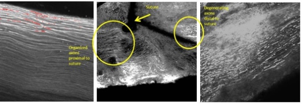 Image 2 - Myelin sheaths 7 days after injury, CARS
 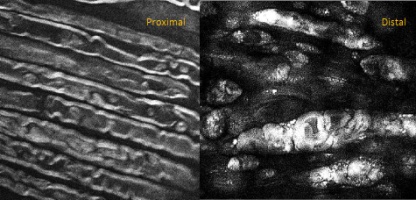 Image 3 ñ Neuromuscular junction, CARS+confocal
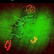
Back to 2014 Annual Meeting Program