
Back to 2014 Annual Meeting Program
Regenerative Peripheral Nerve Interface Viability and Signal Transduction with an Implanted Electrode
Theodore A. Kung, MD1, Nicholas B. Langhals, PhD2, David C. Martin, PhD3, Paul S. Cederna, MD2 and Melanie G. Urbanchek, PhD2
1Department of Surgery, Section of Plastic Surgery, University of Michigan, Ann Arbor, MI, 2Plastic Surgery, University of Michigan, Ann Arbor, MI, 3Materials Science & Engineering, University of Delaware, Newark, DE
Objective: The Regenerative Peripheral Nerve Interface (RPNI) consists of a unit of free muscle that has been neurotized by a transected peripheral nerve. In conjunction with an implanted electrode, the RPNI can facilitate signal transduction from a residual peripheral nerve to a neuroprosthetic limb. Electrodes can be coated with conductive polymer to enhance conductivity. This study examines RPNI viability and signal fidelity in the presence of an implanted electrode. Additionally, this investigation compares the strength of signals recorded from coated and uncoated electrodes. Methods: In a rat model (n=16), the left extensor digitorum longus (EDL) muscle was removed as a nonvascularized free tissue transfer and neurotized by the divided ipsilateral common peroneal nerve. The RPNI was implanted with either a stainless steel pad electrode (SS, n=8) or a pad electrode coated with poly(3,4-ethylenedioxy-thiophene) conductive polymer (PEDOT, n=8). Acellular extracellular matrix secured the electrode against the muscle and isolated the RPNI from surrounding tissues. The contralateral EDL muscle served as Control. Monthly electrophysiological testing was performed using percutaneous stimulation while recording from the indwelling electrode. Results: The free EDL muscle transfers remained healthy and experienced successful revascularization and reinnervation as evidenced by recorded compound muscle action potentials (CMAPs) transduced through the RPNIs and by serial insertional electromyography testing. Histologic examination of the RPNI confirms axonal sprouting, elongation, and synaptogenesis within the RPNI. The conductive polymer coating on the implanted SS electrode resulted in increased electrode recording sensitivity and subsequently increased recorded signal amplitude which was observed throughout the course of the study. Conclusion: The RPNI remains viable over 7 months in the presence of an implanted electrode. Implanted electrodes with and without conductive polymer can be utilized with the RPNI to reliably transduce signals from a transected nerve. Conductive polymer increases the sensitivity of the electrode and results in augmentation of the strength of recorded signals. These findings emphasize the potential of the PRNI as a novel prosthetic interface which may permit dependable control of an artificial limb. 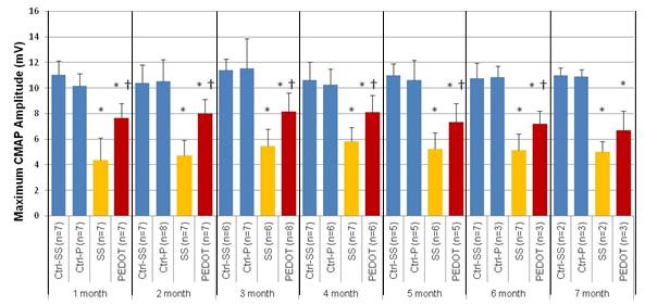
Back to 2014 Annual Meeting Program